|
|
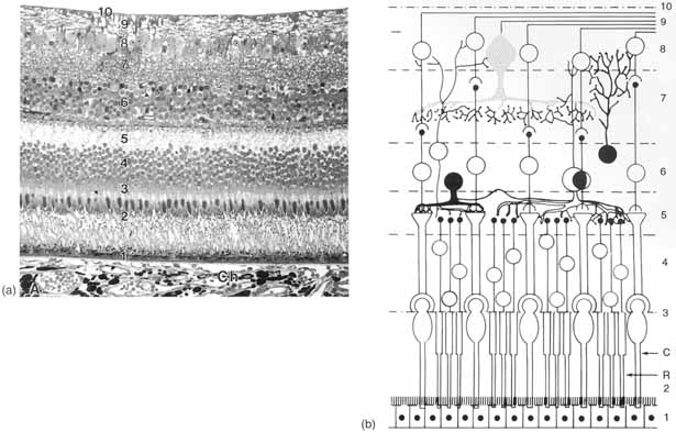
| Fig. 21 Morphological organization of the retina. A. Transverse section of the retina showing pigmented epithelium (1) attached to the sensory retina that consists of photoreceptor layer (2); external limiting membrane (3); outer nuclear layer (4); outer plexiform layer (5); inner nuclear layer (6); inner plexiform layer (7); ganglion cell layer (8); nerve fiber layer (9); and internal limiting membrane (10). Ch, Choroid. Photomicrograph, original magnification ×245. B. Diagrammatic representation of the elements that comprise the retina. 1, Pigment epithelium; 2, photoreceptor layer consisting of rods (R) and cones (C); 3, external limiting membrane; 4, outer nuclear layer; 5, outer plexiform layer; 6, inner nuclear layer; 7, inner plexiform layer; 8, ganglion cell layer; 9, nerve fiber layer; 10, internal limiting membrane. (From Tripathi RC, Tripathi BJ. In: Davson H, ed. The Eye. Academic Press, 1984, with permission) |