|
|
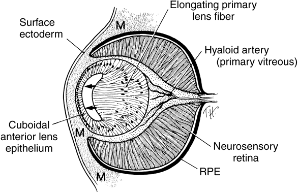
| Fig. 10. Drawing showing formation of the embryonic lens nucleus and primary lens fibers at approximately 6 weeks. Neural crest mesenchyme (M) surrounds the optic cup. The posterior lens epithelial cells (located nearest the developing retina) elongate to form the primary lens fibers. The anterior epithelium remains cuboidal and becomes the anterior epithelium in the adult. The optic fissure is now closed. The hyaloid vessels are seen between the lens and retina. (Cook CS, Sulik KK, Wright KW: Embryology. In Wright KW [ed]: Pediatric Ophthalmology and Strabismus, pp 3–43. St Louis: Mosby, 1995.) |