|
|
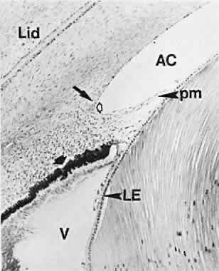
| Fig. 18. Excavation of the anterior chamber (AC) angle in a fetus at 75 mm (3 months) is at a level with the rim of the optic cup, which is well ahead of the lens bow. The corneal endothelium extends to the apex of the angle (hollow arrow). The location of the future trabecular meshwork is indicated by the arrow. On the side toward the lens, the angle is limited by the forward extension of the loosely woven mesenchyme over the optic cup margin. Blood vessels in the recesses of the pigment epithelium (solid arrow) precede its infolding. LE, lens epithelium; pm, pupillary membrane. |