|
|
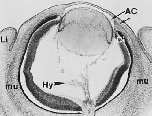
| Fig. 26. General view of the eye at approximately 20 mm (45 days). Ciliary portion of the neural cup (Ci) is relatively undifferentiated and extends to about the level of the lens equator. The mesenchyme around and anteriorly to the margin of the cup shows at least two different degrees of condensation, separated by an interface (arrow). Mesenchymal cells do not yet fill the center of the space between corneal epithelium and endothelium. Anlagen of the extraocular muscles (mu) are recognizable. Upper and lower lids (Li) are undifferentiated skin folds. An anterior chamber (AC) is delineated by the pupillary membrane at the arrow. Major components of the hyaloid vasculature (Hy) are represented. (Smelser GK: Embryology and morphology of the lens. Invest Ophthalmol 4:398, 1965.) |