
|
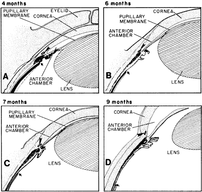
| Fig. 27. Schematic diagram of the developing ciliary body and iris; their relation to the positions of Schlemm's canal and ora serrata. Drawn after sagittal sections of celloidin-embedded eyes. A. At 4 months, a rough triangular anlage of the meridional ciliary muscles fibers (shaded) is present behind the angle recess. Arrow points to the incipient ora serrata behind the most posterior ciliary fold. Ectodermal iris is short and the canal of Schlemm (arrowhead) is behind the bottom of the angle. B. At 6 months, the meridional fibers (shaded) are connected to the scleral spur, behind the angle. Some circular ciliary muscle fibers begin to differentiate and the pars plana and ectodermal iris lengthen. The canal of Schlemm (arrowhead) is mostly behind the level of the deep portion of the angle. The ora serrata is located over the middle portion of the ciliary muscle and is indicated by the arrow. C. At 7 months, one third of the pars plana covers the meridional ciliary muscle and the circular fibers (shaded) are well established. The angle has deepened so that the canal of Schlemm (arrowhead) is at its level. The ora serrata is indicated by the arrow. D. At 9 months, the pars plana lengthens and is over two thirds of the meridional fibers (shaded). The ora serrata is marked by the arrow. The iris is nearly fully developed but still has a thick root. The canal of Schlemm (arrowhead) is in its definitive location anterior to the angle recess. |