|
|
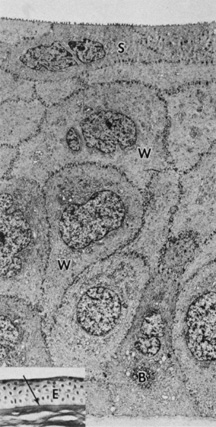
| Fig. 2. Full-thickness electron micrograph of corneal epithelium. Note the cell shape change with depth, the variation of cell membrane interdigitation, and the intracellular differences between cell types. S, apical surface cells; W, wing cells; and B, basal cells. Also note the microvilli seen along the apical membrane of the surface cells (3,620×). Inset: Epithelium (E) overlies a thin, dense basement membrane (arrow) with no discernible laminar appearance (periodic acid-Schiff [PAS] stain, 330×). (Courtesy of Drs. Rodrigues, Waring, Hackett, and Donohoo.) |