|
|
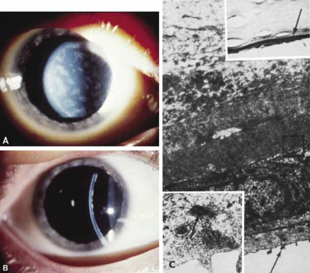
| Fig. 26 Posterior polymorphous dystrophy. A. Classic appearance of geographic vesicular opacities at the level of Descemet's membrane. This type of opacity is not usually diagnosed at birth. B. Another slit view showing these opacities at the level of Descemet's membrane. C. Upper inset shows multiple lamellae of Descemet's membrane with two layers of cells on the posterior cornea (arrow) (Toluidine blue, ×230). Main figure demonstrates abnormal Descemet's membrane consisting of fine lamellae anteriorly and of more homogeneous basement membrane-like material posteriorly studded by 110-nm banded wide-spacing collagen (box). Cells on posterior cornea have epithelial-like morphology with surface microvilli (arrow) (×12,000). Lower inset demonstrates microvilli and desmosome-like intracellular adhesions (arrow) (×56,000). |