
|
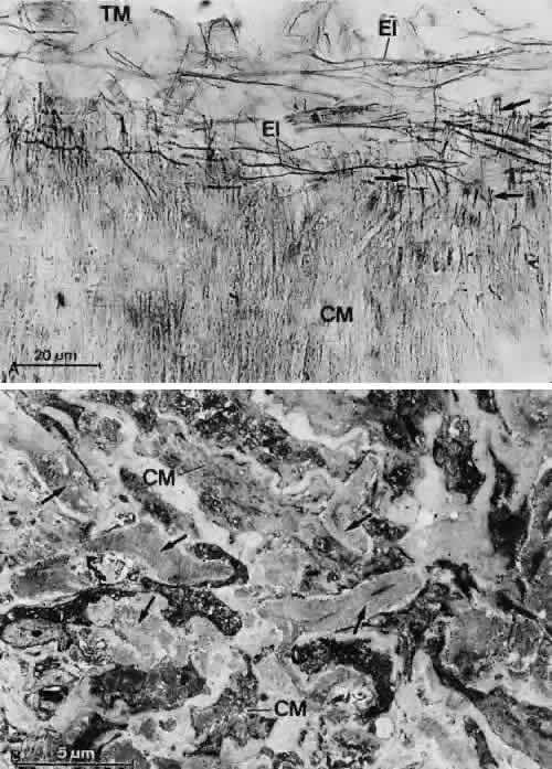
| Fig. 32. Elastic fibers at the anterior ciliary muscle tips. A. Light micrograph of a tangential section through the transition zone of the anterior ciliary muscle tips and the posterior trabecular meshwork (TM) (resorcin fuchsin stain). Note that the network of elastic-like fibers (EI) does not continue posteriorly into the ciliary muscle (CM). Arrows indicate elastic tendons of anterior muscle tips. B. Electron micrograph of a sagittal section through the anterior ciliary muscle tips (chronic simple glaucoma). Within the connective tissue surrounding the muscle fibers (CM), broad strands of elastic-like fibers exist (arrows) that consist of an electron-dense central core and a thick sheath resembling the elastic-like fiber sheath within the trabecular meshwork. |