
|
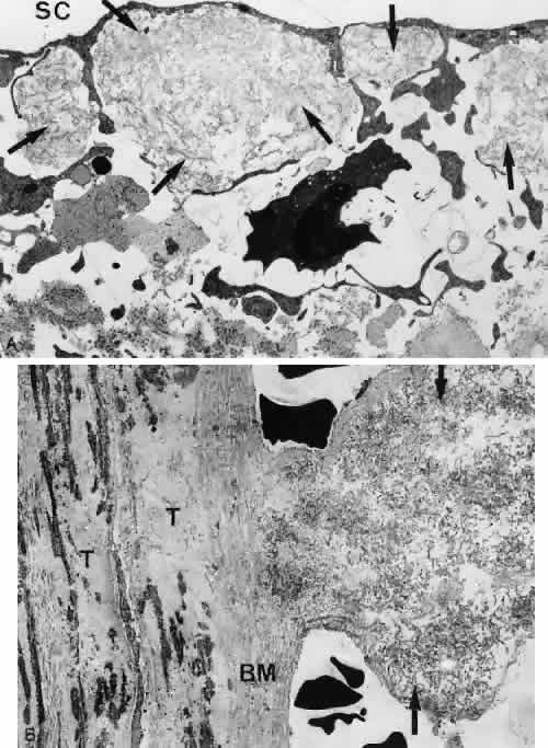
| Fig. 34. Electron micrographs of trabeculectomy specimens taken from cases of pseudoexfoliative glaucoma. A. Inner wall region containing large clusters of exfoliative material (arrows) underneath the endothelial lining of Schlemm's canal (SC) (× 16,560). B. Cross section of a trabecular lamella (T) showing a thickened basement membrane (BM), to which a large cluster of exfoliative material (arrows) is attached (× 30,000). |