
|
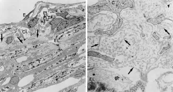
| Fig. 35. Electron micrographs of the trabecular meshwork in a case of corticosteroid glaucoma in a 63-year-old woman. A. Survey figure showing the cribriform layer (CL) and the adjacent corneoscleral lamellae (T). Deposits of extracellular material, typical for corticosteroid glaucoma, are indicated by arrows (× 5,000). E, endothelium of Schlemm's ca-nal; EL, elastic-like fibers; S, subendothelialcells of the cribriform layer. B. Higher magni-fication of the cribriform layer showing thedeposits of extracellular material, characteris-tic for corticosteroid glaucoma (arrows)(× 51,000). C, cells of cribriform layer; EL, elastic-like fiber. |