|
|
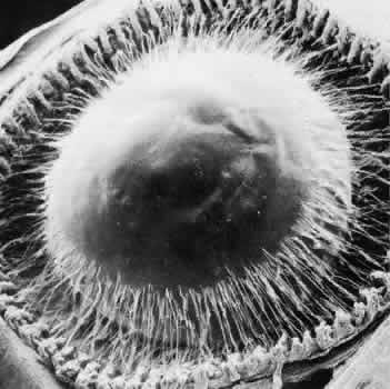
| Fig. 21. Scanning micrograph of the anterior zonular insertion after removal of cornea and iris. The anterior heads of the ciliary processes are free of zonules, leaving an empty V as the zonular bundles enter on each side of the processes. Lens is 25% smaller than normal owing to processing shrinkage (× 25). |