
|
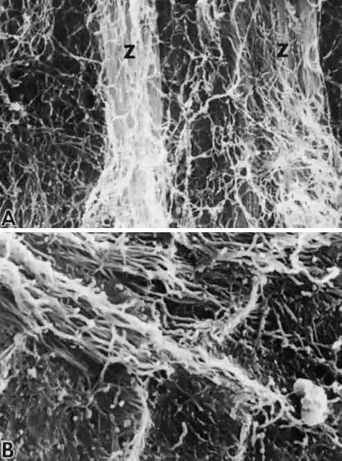
| Fig. 34. Insertion of the posterior zonular fibers. A. A thick layer of looser fibrils covers the inserting zonular fibrils (Z) (SEM, × 9,200). B. When exposed, the deep insertion shows the usual blending of zonular fibrils into the fibrogranular matrix of the superficial capsule (SEM, × 15,000). (Streeten BW, Pulaski JP: Posterior zonules and lens extraction. Arch Ophthalmol 96:132, 1978. Copyright 1978, American Medical Association) |