|
|
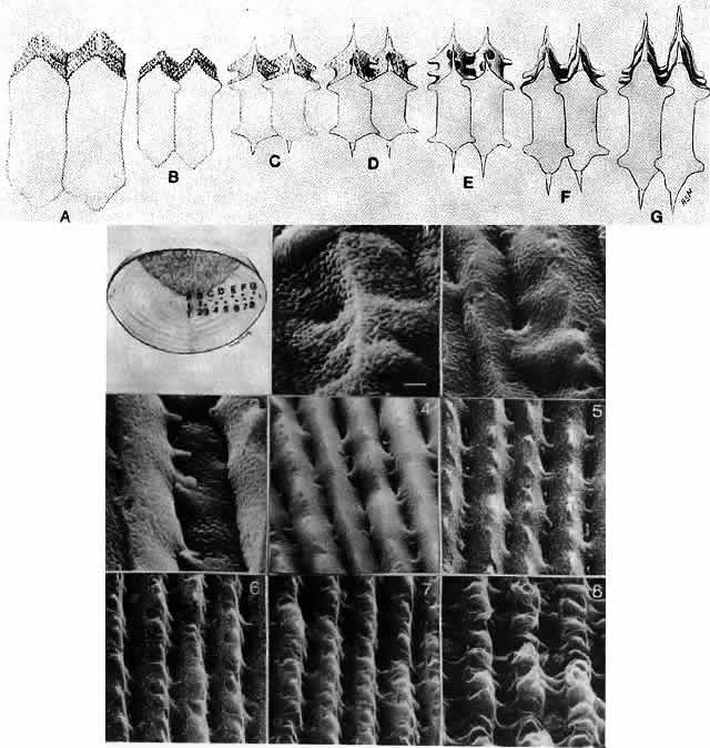
| Fig. 25. The differences in lens fiber morphology from various regions of the rabbit lens. The uppermost schematic and the scanning electron micrographs are keyed to the labeled sagittal diagram of the lens. The numbers correspond to those shown on the eight scanning electron micrographs. The fibers in each case are oriented so that the narrow side of the hexagon is exposed. The scale bar in the first plate represents 1 μm. (Harding CV, Susan S, Murphy H: Scanning electron microscopy of the adult rabbit lens. Ophthalmol Res 9:443, 1976) |