|
|
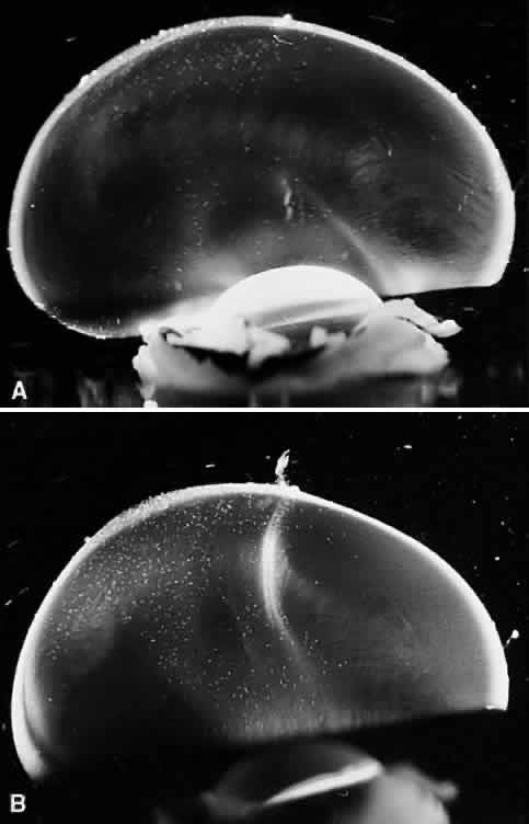
| Fig. 9. Vitreous structure in the human embryo. These specimens from a 33-week-old human embryo have had the sclera, choroid, and retina dissected off the vitreous body, which is still attached to the anterior segment (bottoms of photos). A: The central vitreous is relatively clear, although there is more light scattering than in the postnatal period, most likely because of the relative paucity of vitreous HA at this stage of development. The vitreous cortex has considerable light scattering because of the high density of collagen fibrils in the peripheral shell of the vitreous body. B: Cloquet's canal is seen coursing in an antero-posterior orientation, arising from the posterior aspect of the lens and orienting toward the optic disc. (From Sebag J. Age-related changes in human vitreous structure. Graefes Arch Clin Exp Ophthalmol 225:89, 1987, with permission) |