|
|
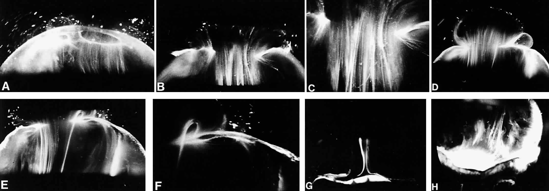
| Fig. 12. Adult human vitreous morphology. All photographs were taken from human eyes after dissection of the sclera, choroid, and retina, with the vitreous still attached to the anterior segment. A slit-lamp beam shown from the side illuminated a plane through the specimens, and photographs were taken at a 90-degree angle to this plane, thereby maximizing the Tyndall effect. The anterior segment is below and the posterior pole is above in all photographs. A: Posterior vitreous in the left eye of a 52-year-old man. The corpus vitreus is enclosed by the vitreous cortex. There is a hole in the prepapillary (small hole to the left) and a dehiscence in the premacular vitreous cortex. Vitreous fibers are oriented toward the premacular vitreous cortex. B: Posterior vitreous in a 57-year-old man. A large bundle of prominent fibers is seen coursing anteroposteriorly to exit by the premacular dehiscence in the vitreous cortex. C: Same as (B) at higher magnification. D: Posterior vitreous in the right eye of a 53-year-old woman. There is extrusion of the central vitreous by the prepapillary hole (to the right) in the vitreous cortex and the premacular (left) vitreous cortex. Fibers course out into the retrocortical (preretinal) space. E: Same specimen as (D) at a different level of horizontal optical sectioning. A large fiber courses posteriorly from the central vitreous and inserts into the posterior vitreous cortex at the rim of the premacular dehiscence in the cortex. F: Same as (E) at higher magnification. The large fiber has a curvilinear appearance because of traction by the vitreous extruding out into the retrocortical space. Because of its attachment to the vitreous cortex, the fiber arcs back to its point of insertion. G: Anterior and central vitreous in a 33-year-old woman. The posterior aspect of the lens is seen below. Cloquet's canal is seen forming the retrolental space of Berger. H: Anterior and peripheral vitreous in a 57-year-old man. The specimen is tilted forward to show the posterior aspect of the lens and the peripheral anterior vitreous. Behind and to the right of the lens there are fibers coursing anteroposteriorly to insert into the vitreous base. Within the vitreous base, these fibers splay out to insert anterior and posterior to the ora serrata. (A, E, and F from Sebag J, Balazs EA. Pathogenesis of C.M.E.–an anatomic consideration of vitreo-retinal adhesions. Surv Ophthalmol 28:493, 1984, with permission; B and C from Sebag J, Balazs EA. Morphology and ultrastructure of human vitreous fibers. Invest Ophthalmol Vis Sci 30:187, 1989, with permission) |