|
|
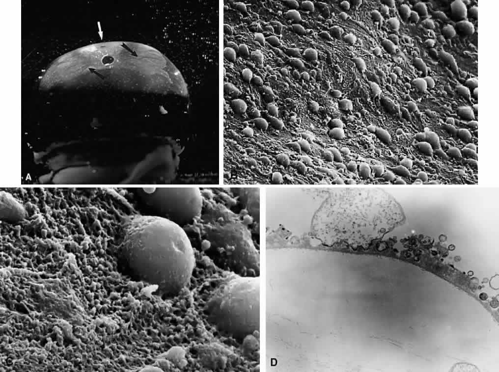
| Fig. 16. Vitreoretinal interface in youth. A: Dark-field microscopy of the posterior vitreous in a 14-year-old boy. The sclera, choroid, and retina were dissected off the corpus vitreus, which remains attached to the anterior segment. In contrast to adults, there is an extra layer of tissue that remained adherent to the posterior vitreous cortex when the retina was dissected off. The white arrow indicates the location of the fovea. The circular structure below this location is the prepapillary hole in the posterior vitreous cortex. Emanating from this hole are linear, branching structures (black arrows) that correspond to the location of the retinal vessels. B: Scanning electron microscopy of the tissue described in (A) demonstrates many round structures adherent to the posterior aspect of the tissue. Bar = 10 μm. C: Higher magnification showing the attachment of one of these round structures. There appears to be an indentation or hole on the posterior aspect of this structure. Bar = 1 μm. D: Transmission electron microscopy of this specimen identifies this tissue as the internal limiting lamina (ILL) of the retina attached to the posterior vitreous cortex. The round structures are identified as the inner portion of Müller cells that remained adherent to the posterior aspect of the ILL, with a hole on the posterior aspect of the inner portion of the Müller cell where it was torn away from the rest of the cell body (×20,800). (From Sebag J. Age-related differences in the human vitreoretinal interface. Arch Ophthalmol 109:966, 1991, with permission) |