|
|
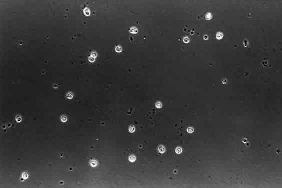
| Fig. 19. Human hyalocytes in situ. Phase-contrast microscopy of flat-mount preparation of posterior vitreous cortex from the eye of an 11-year-old girl obtained at autopsy (courtesy of New England Eye Bank, Boston, MA). No stains or dyes were used in this preparation. Mononuclear cells are distributed in a single layer within the vitreous cortex (7times;115). |