|
|
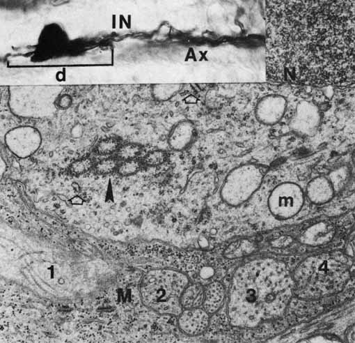
| Fig. 28 Portion of a horizontal cell of a region with some of Kolmer's crystalloid, sectioned slightly diagonally (arrowhead). Other cytoplasmic components are microtubules, vesicles, vesicular and tubular smooth endoplasmic reticulum, granular endoplasmic reticulum (open arrowheads), polysomes, and mitochondria (m) whose loss of detail is an artifact. Kolmer's crystalloid represents a more structured rough endoplasmic reticulum. The portion of the Müller cell (M) on the bottom has glycogen granules and surrounds longitudinally (1) and transversely (2, 3, 4) cut axons and dendrites of the first synaptic area. N, nucleus (original magnification ×24,000). Inset. Photomicrograph of a Golgi-impregnated horizontal cell. Total magnification is ×1120. Ax, axon through only part of its length; d, portion of dendritic spread that is visible in this section; IN, part of inner nuclear layer. |