
|
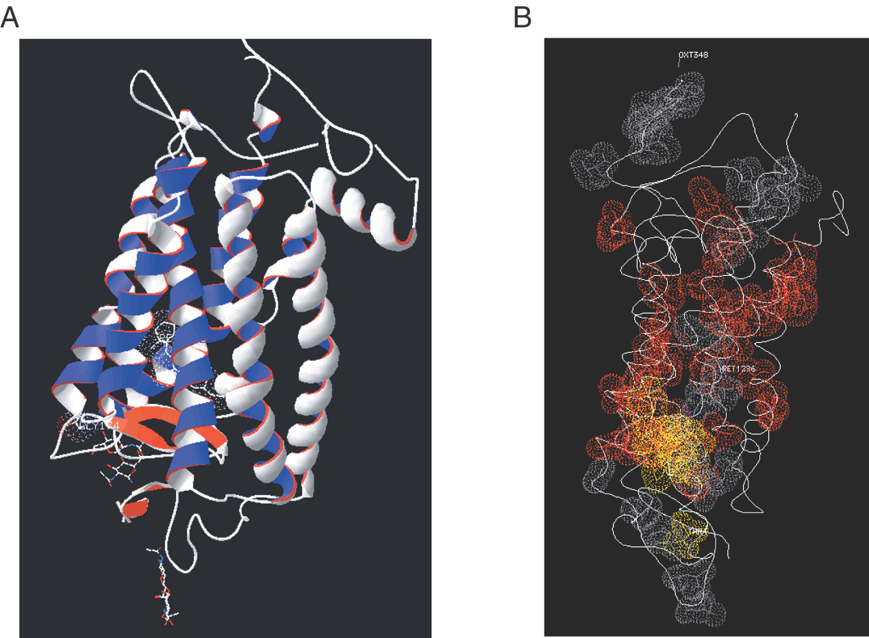
| Fig. 18. Structure of rhodopsin and locations of missense mutations. Panel A. A depiction of the 3D structure of bovine rhodopsin based on X-ray diffraction of rhodopsin crystals, and Protein Database (PDB) entry 1L9H. Transmembrane helices are represented by the gray and blue ribbons with a red edge. Beta strands are illustrated in orange and yellow-tan. The C-terminal end of the strands is identified by the arrow. The four strands constitute the plug region on the extracellular (luminal) side of the protein. Panel B. A quicktime video on the accompanying CD illustrates the positions of known missense mutations causing autosomal dominant retinitis pigmentosa (ADRP) and congenital stationary night blindness (CSNB) in the 3D structure of rhodopsin. In this version, panel B represents the first image of the video. The opsin coordinates are taken from PDB entry 1L9H, which provides resolution refined to 2.6 Å. In the video version, the reader should be able to locate several domains devoid of mutations (illustrated by ribbons lacking spheres) and other regions that contain many mutations (represented by the spheres). The luminal side shows many mutations in the plug and near the retinoid-binding site. The positions of residues mutated in ADRP and CSNB are illustrated by surface dots that represent van der Waal's radii. Normal amino acid residues are shown. |