
|
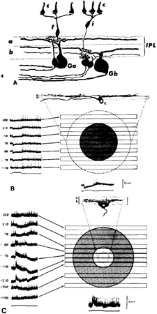
| Fig. 17. A. Organization of cone bipolar cells and ganglion cells in the inner plexiform layer (IPL) of the cat retina. Flat cone bipolar cells (f) have axon terminals that end in sublamina a and contact the dendrites of Ga-type ganglion cells. Invaginating cone bipolar cells (i) have axon terminals that ramify at a more proximal level of the IPL in sublamina b, where they contact Gb-type ganglion cells. Ga cells are off-center and Gb cells are on-center ganglion cells. c, cones. B. Receptive field properties and morphology of an intracellularly stained off-center ganglion cell. Note the dendritic arborization in sublamina a. At left are shown responses to slits of light positioned at different levels of the receptive field. They show that a hyperpolarization (inhibitory postsynaptic potential) is elicited when the slit is positioned between 210 μm and -70 μm (shaded circle). The dotted circle gives the dendritic field of the cell as 490 μm. The response to a diffuse stimulus is shown at bottom. (Stimulus width, 50 μm; duration, 542 msec; wavelength, 441 nm; intensity, 2.7 log quanta μm-2 sec-1). c, cone. C. Concentrically organized on-center ganglion cell of the rat retina. The dendritic arbor of this cell is confined to sublamina b of the IPL. The membrane depolarization (excitatory postsynaptic potential) is elicited by light stimuli falling between 0 and 210 μm (open circle). The dotted circle indicates the subtense of the cell's dendritic arbor. The shaded circle indicates the spatial extent of the inhibitory surround. (Stimulus width, 100 μm; duration, 560 msec; wavelength, 647 nm; intensity, 4.6 log quanta μm-2 sec-1) (Nelson R, Famiglietti EV Jr, Kolb H: Intracellular staining reveals different levels of stratification for on- and off-center ganglion cells in cat retina. J Neurophysiol 41:472, 1978) |