
|
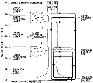
| Fig. 22. Frog retina. A simplified diagram illustrating the postulated role of the Muller fiber in the generation of the ERG b wave. Light evokes an increase in the extracellular potassium concentration in both plexiform layers. Current flows into the Muller fiber at these regions and exits mainly at the Muller fiber vitreal end foot. The sink/source profile of the Muller fiber current plays a major role in the generation of the b wave. (Newman EA: Current-source density analysis of the b-wave of frog retina. J Neurophysiol 43:1355, 1980) |