|
|
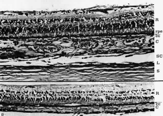
| Fig. 3. Choroid. A. The layers of medium and large choroidal vessels thicken the choroid in the submacular region. (PAS, × 125) B. Peripheral choroid in same patient. Retina and choroid are thinner. Choriocapillaris occupies about one third of choroid, a greater proportion than in posterior pole. The depth of capillaries in periphery and posterior pole is about the same. Outer choroid (C) is reduced to thin layer of arteries and veins. R = retina; rpe = retinal pigment epithelium; arrow in margin = Bruch's membrane; cc = choriocapillaris; SC = suprachoroidal space; L = lamina fusca; S = sclera; single arrow = arteries; double arrow = veins. (PAS, × 125) |