|
|
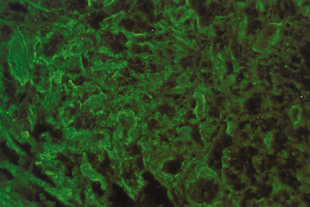
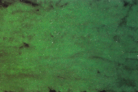
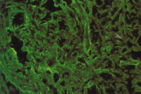
| Fig. 15. Immunofluorescence microscopy of human adult sclera using anticollagen type I (A), anticollagen type II (B), and anticollagen type III (C), antibodies (100×). Collagens type I and III stain intensely in sclera whereas collagen type II is absent. |