|
|
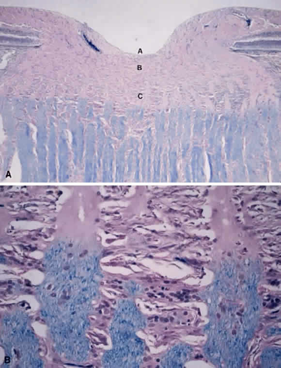
| Fig. 2. A. Histology of the optic nerve head. Longitudinal section: (A) the surface nerve fiber layer and the physiologic cup. (B) Prelaminar region. (C) Lamina cribrosa region. The nonmyelinated axons in the optic nerve head are not stained by Luxol fast blue, whereas the myelinated axons behind the lamina cribrosa are stained. Luxol fast blue, × 40. B. High magnification demonstrates the transmission of the nonmyelinated and myelinated axons, × 400. |