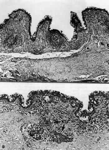
Fig. 7.
A.
Inferior fornix showing papillary projection (
P
).
B.
Inferior fornix demonstrating Krause's glands (
KG
). (
A,
× 40;
B,
× 80)