|
|
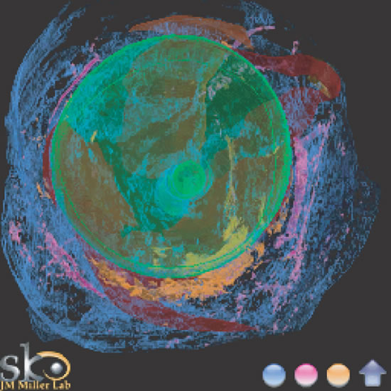
| Fig. 11. Extraocular tissue architecture, anterior view. Serial sections of human orbit, stacked and reassembled by computer. View from front. Green, sclera; Red, muscle; Pink, smooth muscle; Blue, collagen; Orange, elastin. Note heavy elastin and smooth muscle concentration along the inferior half of the globe, constituting part of Lockwood's ligament. (Miller JM, Demer JL, Poukens V et al:Extraocular Tissue Architecture H7, privately published Quicktime VR scene. © 2002 Joel M. Miller.) |