|
|
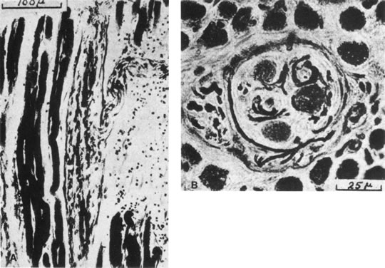
| Fig. 27. Longitudinal sections (A) and cross sections (B) of human extraocular muscle spindles. The torpedo-shaped capsule is delicate and is separated from the intrafusal fibers by a periaxial space. The longitudinal section shows the point of entry of a nerve trunk and at least two intrafusal muscle fibers, which are smaller in diameter than the extrafusal fiber. A capillary runs within the capsule along its left side. The cross section shows three intrafusal fibers and several nerve fibers and capillaries. (Cooper S, Daniel PM: Muscle spindles in human extrinsic eye muscles. Brain 72:1, 1949.) |