
|
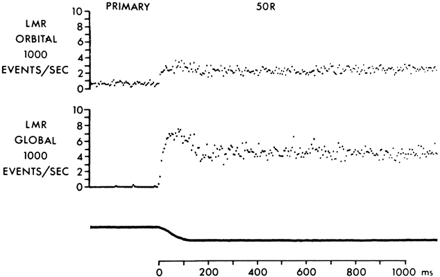
| Fig. 35. Electromyogram of single-unit activity in the human left medial rectus muscle during a 50-degree rightward saccade. The top tracing shows the orbital layer; the bottom tracing shows the global layer, Note that the orbital layer shows a step of innervation, appropriate for positioning the pulley against a spring load. The global layer shows both a pulse and a step, which is required to overcome inertial mass and tissue viscosity prior to giving a holding force appropriate for the new gaze direction. (Collins CC: The human oculomotor control system. In Lennerstrand G, Bach-y-Rita P (eds): Basic Mechanisms of Ocular Motility and Their Clinical Implications, pp 145–189. New York, Pergamon Press, 1975.) |