
|
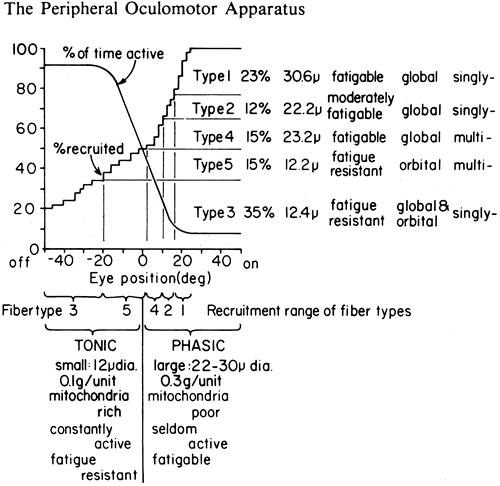
| Fig. 36. Hypothetical functional arrangement of eye muscle fiber types. The type nomenclature and data are those of Alvarado and Van Horn.49 The data to the right of the type number are the percentages of total fibers for each type, average diameter, fatigability, location in orbital or global layer, and single or multiple innervation. One curve plots the percentage of fibers recruited at any gaze angle, the other the percentage of time the fiber is active. The fatigue-resistant fibers are constantly active out of the field of action of the muscle and are termed “tonic” here. The fatigable fibers (“phasic”) are recruited in the field of action of a muscle. The multiply innervated tonic fibers are recruited immediately on either side of primary position. (Robinson DA: The functional behavior of the peripheral ocular motor apparatus: a review. In Kommerell G (ed): Disorders of Ocular Motility: Neurophysiological and Clinical Aspects, pp. 43–61. Presented before the Symposium of Deutschen Ophthalmologischen Gesellschaft, Freiburg, April 1977. Munich, Bergman, 1978. |