|
|
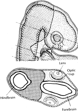
| Fig. 1. Parasagittal and axial sections through the head of an embryo show the eventual ventral location of the migrated neural crest cells (dotted area) in relation to the paraxial mesoderm (crosshatched area) around the hindbrain. (Johnston MC, Bhakdinaronk A, Reid YC: An expanded role of the neural crest in oral and pharyngeal development. In Bosma JF [ed]: The Fourth Symposium on Oral Sensation and Perception, pp 37–52. HEW Pub No 73–546. Bethesda, National Institutes of Health, 1973) |