|
|
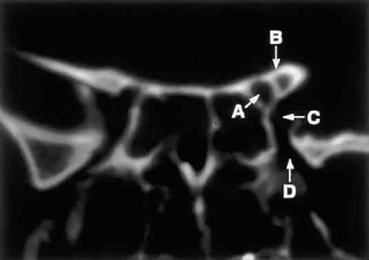
| Fig. 8. Coronal CT image (bone window) of the orbital apex in a patient with facial trauma. Note the position of the posterior orbital foramina. The optic canal (A) is always seen in conjunction with the laterally adjacent anterior clinoid process (B) on both axial and coronal views. Slightly lower, the superior orbital fissure (C) communicates with the CS, found directly behind it. The inferior orbital fissure (D) provides communication through the orbital floor with the pterygopalatine fossa. |