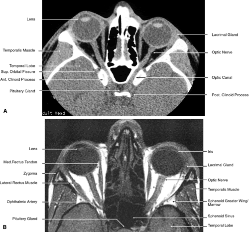
Fig. 23.
Axial images at the level of midorbit.
A.
Computed tomography scan.
B.
T1-weighted magnetic resonance imaging.