Chapter 11
Iris
D. HUNTER CHERWEK, HANS E. GROSSNIKLAUS and AMY K. HUTCHINSON
Main Menu   Table Of Contents

Search

INTRODUCTION
EMBRYOLOGY
GROSS ANATOMY
ANTERIOR IRIS SURFACE
POSTERIOR IRIS SURFACE
VASCULAR SUPPLY
NERVE SUPPLY
HISTOLOGY AND ELECTRON MICROSCOPY
ANTERIOR BORDER LAYER
STROMA
BLOOD VESSELS AND NERVES
SPHINCTER MUSCLE
DILATOR MUSCLE AND ANTERIOR PIGMENT EPITHELIUM
POSTERIOR PIGMENT EPITHELIUM
REFERENCES

INTRODUCTION
The iris functions as a diaphragm within the eye, separating the anterior and posterior chambers of the eye. The structure is named after Iris, the Greek goddess of the rainbow, and provides us with our characteristic eye color. Its central opening, the pupil, is the aperture that regulates the amount of light entering the eye. Although seemingly simple in structure, iris function is complex and affects image clarity. Normal and pathologic states of the efferent and afferent visual pathways are clinically evaluated via pupillary function. The iris is the most anterior part of the uveal tract and can be involved in many ocular and systemic diseases. This chapter discusses the embryology, gross anatomy, histology, and ultrastructural features of the iris.
Back to Top
EMBRYOLOGY
Historically, the embryology of the iris was based on the classical theory of development, which described three germ layers in the developing embryo: endoderm, mesoderm, and ectoderm.1–3 It is now known that the majority of the ocular mesenchyme is derived from neural crest cells.4–7 Neural crest cells are defined as those neuroectodermal cells that proliferate from the crest of the neural folds before the formation of the neural tube. Recently, certain genetic transcription factors, such as PAX6, have been shown to play a key role in orchestrating the formation of the developing tissues in the anterior segment, and mutations of such genes have been implicated in certain diseases of the iris, including aniridia.8–9

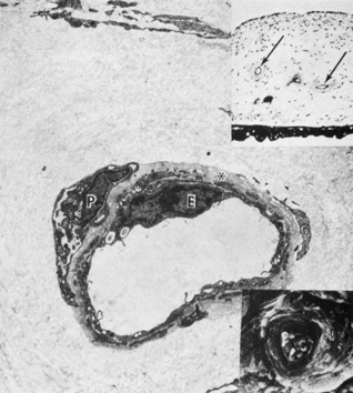
Embryologically, the structure of the iris can be divided into two different developmental layers: iris pigment epithelium (IPE) of neuroectodermal origin and iris stroma of neural crest origin. The cells of the IPE arise from mesoderm, and the stromal blood vessels begin as penetrating branches from the developing vascular network of the ciliary body (Fig. 1). Although data regarding the origin of both iris muscles (sphincter and dilator) are incomplete, they likely arise from neuroectoderm.4,6

Fig. 1. As demonstrated, the iris is derived from both neural crest cells and the optic cup. The Anterior Epithelial Layer (AEL) and Posterior Epithelial Layer (PEL) are derived from the optic cup and are oriented in an apex-to-apex pattern with basement membranes (BM). When the five layers of the iris are seen in cross section, one can see that the Anterior Border Layer (ABL) and stroma (colored in blue) are derived from the neural crest cells and the iris muscles; AEL and PEL (shaded in orange) come from the optic cup.

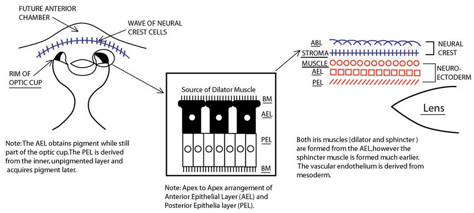
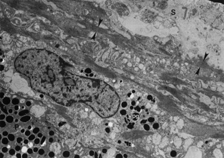
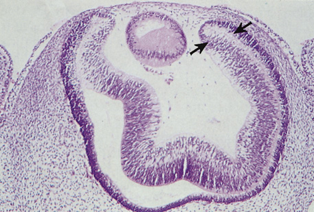
The embryologic precursors of the iris are first visible at six weeks gestational age. The neural ectodermal derivatives (iris epithelium and muscle) arise from the anterior rim of the optic cup. At about six weeks gestational age, these cells begin to proliferate rapidly and extend anteriorly.10 During development, an apparent dilation, called the marginal sinus of von Szily, can be seen at the tip of the optic cup between the two layers of epithelium. Although this sinus is now thought to represent an artifact, the two layers of neuroepithelium become functionally divided into the anterior and posterior pigment epithelium (Fig. 2).11 Embryologically, the anterior epithelium becomes pigmented while still part of the optic cup; however, the pigmentation of the posterior epithelium (derived from the inner, unpigmented layer of the optic cup) occurs later, proceeding toward the ciliary region, and is completed by seven months gestation.12

Fig. 2. The two layers of the neuroepithelium of the optic cup (arrows) become the anterior and posterior iris pigment epithelium, and the iris stroma is derived from neural crest tissue (hematoxylin-eosin, ×63).

Differentiation of the iris stromal elements begins slightly earlier than iris neuroepithelial growth and proceeds as a wave of mesenchymal migration at about six weeks' gestation. This forms the anlage of iris stroma, a continuous papillary membrane, and the tunica vasculosa lentis (a transient vascular network that supplies the developing lens). Iris stromal pigmentation does not occur until after 24 weeks. The developing iris lacks a pupil because the original stroma is a continuous sheet of tissue with epithelial cells growing inward, toward the center of the iris.4,11

The vascular framework of the iris also begins at six weeks as a blind axial outgrowth from the annular vessels in the mesenchyme associated with the rim of the optic cup and forms the anterior part of the tunica vasculosa lentis (lamina iridopupillaris). By the third month, branches of the nasal and the temporal long posterior ciliary arteries unite with peripheral vessels of the lamina iridopupillaris to form the major arterial circle. The pupil is created by cellular and vascular remodeling of the iridopupillary membrane during the fifth month of gestation.4,11

The development of the iris sphincter muscle first appears as basal infoldings in the cells of the anterior layer of neuroepithelium and can be seen by light microscopy at about eleven weeks. By the fifth month, myofibrils begin to be synthesized by the developing tissue; the muscle comes to lie free in the posterior mesenchymal layer by the eighth month of gestation. The formation of the dilator muscle fibers occurs later and is first identified in the sixth month as fine fibrils in the anterior iris epithelium.

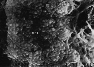
The iris is near its adult size at birth. The most dynamic changes occur in the first few postnatal years and involve the color of the iris and the extracellular matrix. Embryologically, the anterior epithelium becomes pigmented while still part of the optic cup; however, the pigmentation of the posterior epithelium (derived from the inner, unpigmented layer of the optic cup) occurs later, proceeding toward the ciliary region, and is completed by seven months' gestation. Postnatal changes in iris color occur by the gradual accumulation of melanin in the stroma and anterior border layer as discussed later.

Back to Top
GROSS ANATOMY
The iris measures approximately 12 mm in diameter and about 37 to 38 mm in circumference. It is thickest at its central portion (the collarette) and thinnest near the ciliary body's insertion point (the root, the site most prone to tears from trauma). The pupil is decentered somewhat nasally and inferiorly to the central cornea with the temporal iris being broader than the nasal iris. Anteriorly, the iris is bathed by aqueous humor. Posteriorly, the central portion of the iris abuts the lens, which displaces the iris somewhat anteriorly and results in its characteristic conical configuration. Physical analyses of the factors that determine the contour of the iris are available.13,14 Peripherally, the posterior iris is bathed by the aqueous in the posterior chamber.

The color of the iris is determined by three main variables: (1) the density and structure of the iris stroma, (2) the pigment epithelium, and (3) the pigment content (granules) within the melanocytes of the iris stroma.15 Eagle suggests that the color of the iris is determined not by the number of melanocytes in the iris stroma, as previously believed, but rather by the character of the melanin granules within superficial stromal melanocytes.16 Therefore, it should be noted that the number and distribution of melanocytes do not vary between the different color irides. A brown iris contains more pigment on its anterior surface and within its stroma than a blue iris. The color of the blue irides results from stromal absorption of the long wavelengths, allowing the shorter blue wavelengths to be reflected back to the observer because the stroma is relatively free of pigment.16–18 Tyrosinase-negative albino irides have been shown to have stromal melanocytes containing mature type IV melanosomes19; however, the characteristic pink color of the iris occurs because the only coloration present is from the background red reflex of the retina and from blood within the iris vessels. More recently, investigators have begun to isolate genetic markers that are associated with certain iris colors; the complex genetic components of iris pigmentation are only beginning to be understood.20

The recent use of topical prostaglandin therapy in the treatment of glaucoma has shown that the color of the iris may also change in some adult patients, which may provide insight into the possible cellular pathways that control iris melanocytes. There are several proposed explanations: (1) the drugs may cause the melanocytes to proliferate; (2) prostaglandins may act directly or through a second messenger on melanocytes, causing either increased tyrosinase activity or decreased breakdown of melanosomes; (3) prostaglandins may cause migration of stromal melanocytes; or (4) these drugs may influence the responsiveness of melanocytes to neuronal stimuli.21–23

Back to Top
ANTERIOR IRIS SURFACE
The collarette of the iris is located about 1.5 mm from the pupillary margin, represents the thickest portion of the iris (0.6 mm), and divides the iris into two zones: the pupillary zone and the ciliary zone (Fig. 3). The pupillary zone begins at the margin of the pupil, known as the pupillary ruff, which represents the anterior termination of the iris pigment epithelium. Numerous crenations are seen in this area and vary with pupillary size. Thick branching trabeculae are most prominently seen in the pupillary and collarette regions. These bands are radially oriented and enclose depressions in the iris surface. Fuchs' crypts are adjacent to the collarette on both its pupillary and ciliary sides. These crypts are focal defects in the anterior layer of the iris stroma, which arise from agenesis, or more likely, atrophy of this layer coincident with resorption of the embryonic pupillary membrane.16 The anterior surface of the ciliary portion has been divided into three areas: an inner smooth area, a middle furrowed area, and a marginal cribiform area (which is visible only by gonioscopy). The entire ciliary region is characterized by a pattern of radial ridges formed by underlying stromal blood vessels.17

Fig. 3. The anterior iris surface features the inner pupillary zone and outer ciliary zone. Crypts (arrowhead) and marginal furrows (arrow) are present.

Back to Top
POSTERIOR IRIS SURFACE
The posterior surface of the iris is much smoother and more uniform than its anterior counterpart (Fig. 4). The posterior surface of the iris contains two layers of epithelial cells, the anterior and posterior iris epithelia. The surface is smooth but broken by relatively shallow furrows. Two types of radial folds are identified: (1) contraction folds of Schwalbe, which are small sulci that run for a distance of 1 mm, bending over the pupillary aperture and forming the crenations on its anterior surface; and (2) structural folds of Schwalbe, which begin about 1.5 mm from the pupillary border and extend between the ciliary processes. Circular contraction furrows are also present.17

Fig. 4. The posterior surface of the iris is smoother than the anterior surface. Radial folds are present.

Back to Top
VASCULAR SUPPLY
Blood supply to the iris originates in the major arterial circle, which lies in the stroma of the ciliary body near the iris root. The vessels have a slight corkscrew shape to accommodate iris dilation and contraction. Branches travel in the anterior iris stroma, extending toward the collarette where they form an incomplete minor arterial circle. Numerous vessels from the minor arterial circle form vascular arcades that course toward the pupillary margin to supply the sphincter muscle and also travel along the dilator muscle toward the iris root. The endothelial cells along the iris vessels are connected by tight junctions and, thus, are impermeable to macromolecules, except during periods of inflammation, as seen with uveitis.24 Venous channels follow a similar course as the arterial system of the iris; however, there is no venous equivalent of the major arterial circlet, and the venous system of the iris eventually drains into the vortex system and ciliary plexus.7
Back to Top
NERVE SUPPLY
The nerve supply of the iris is composed of the long and short ciliary nerves. The long ciliary nerves originate from the first branch of the trigeminal nerve and contain the sensory fibers of the iris. Sensory nerves are distributed diffusely in the stroma, terminating as unmyelinated fibers. Postganglionic sympathetic fibers from the superior cervical sympathetic ganglion also travel with the long ciliary nerves and innervate the dilator pupillae muscle and control vasomotor function of the blood vessels. The short ciliary nerves arise from the ciliary ganglion and contain postganglionic parasympathetic fibers (originating from the oculomotor nerve) and innervate the sphincter pupillae muscle.7 A report describing the receptorial distribution of human iris musculature has shown alpha-exciting and beta-2-relaxing receptors, as well as the absence of cholinergic, serotonergic, dopaminergic, and histaminergic receptors on the iris dilating muscle. On the papillary sphincter muscle, muscarinic cholinergic receptors are present and adrenergic receptors are absent.25 The nerves of the iris are partially encased by thin, cytoplasmic processes of the stromal fibroblasts.
Back to Top
HISTOLOGY AND ELECTRON MICROSCOPY
The iris can be divided into five layers: the anterior border layer, the stroma, the muscle layer, the anterior pigment epithelium, and the posterior pigment epithelium (Fig. 5). The histology and electron microscopic features of each are discussed in the paragraphs that follow.

Fig. 5. The histology of the iris may be divided into five layers: anterior border (A), stroma (S), muscle (M) , anterior pigment epithelium (AP) , and osterior pigment epithelium (PE) (hematoxylin-eosin, ×63).

Back to Top
ANTERIOR BORDER LAYER
During embryologic development, the anterior surface of the iris is covered with a continuous layer of endothelium, which disappears at or soon after birth.7,26–28 Thus, the anterior border layer is a modification of the stroma that is composed of a relatively dense meshwork of melanocytes and fibroblasts with associated collagen. This meshwork of cells does not form a continuous, impermeable sheet that overlies the anterior iris surface and allows aqueous humor to pass freely into the iris stroma. These interrupted gaps or defects within the anterior border layer (i.e., Fuchs crypts, as described earlier) provide each individual patient with a unique topographic “fingerprint,” because iris patterns vary from person to person. Therefore, the anterior border layer filters aqueous into the stroma and begins to filter ambient light as an early light-absorbing layer of the iris.29,30

Eagle16 found relatively constant numbers of melanocytes in the anterior border layer of all irides, regardless of iris color. However, he found that darker irides had more abundant and larger melanin granules, and that the cytoplasmic volume of melanocytes was increased (due to an increase in pigment volume) in darker irides. Eagle also studied features of the anterior border layer of different color irides extensively using a scanning electron microscopic. Lighter irides have delicate processes, and their anterior borders have an intricate plexiform character. Darker irides have fewer processes, which are stout and less dendritic than in lighter irides. Additionally, scattered round processes packed with melanin pigment are sometimes seen in darker irides. The anterior border layer is fairly thick near the pupillary border where it is firmly attached to the two layers of pigment epithelium that form the pupillary ruff (Fig. 6). The thickness of the anterior border layer varies over the surface of the iris. Anterior border cells are generally sparse or absent overlying iris crypts and are dense over contraction furrows, over trabeculae, and in darker irides. The anterior border layer ends at the iris root but may extend in a spokelike fashion to Schwalbe's line as iris processes. Iris freckles are common and are formed by accumulations of pigmented melanocytes present on the anterior iris surface (Figs. 7 and 8).29

Fig.6. Pupillary border of the iris showing slight physiologic ectropion uveae (asterisks). The sphincter muscle is indicated by S (scanning electron microscopy, ×240). Inset of pupillary region shows the sphincter muscle (S) and pigment epithelium (P) (Toluidine blue, ×300).

Fig. 7. The anterior border layer (ABL) of the iris is densely pigmented in an area of freckles. The uveal melanocytes display a dendritic pattern (hematoxylin-eosin, ×220).

Fig. 8. An iris freckle composed of melanocytes (MEL) with prominent pigment granules is present on the anterior iris surface (scanning electron microscopy, ×5400).

Fibroblasts are prominent on the iris surface. Transmission electron microscopic examination of these fibroblasts reveals abundant mitochondria, rough endoplasmic reticula, free ribosomes, and bundles of filaments (Fig. 9). Some fibroblasts have basal bodies in their cytoplasm, with associated cilia projecting into the anterior chamber (Figs. 10 and 11). Melanocytes are most often found just beneath fibroblasts and contain mitochondria, smooth and rough endoplasmic reticula, free ribosomes, and melanin granules in various stages of development (Fig. 12). The blood vessels and nerve fibers of the anterior border layer are similar to those in the deeper stroma and are discussed later.29

Fig. 9. The anterior border of the iris is composed of a discontinuous layer of fibroblasts (F) and interspersed collagen fibrils (arrows) (×13,800). Inset shows fibroblasts and a meshwork of collagen connecting fibroblasts (×40,500).

Fig. 10. Microvilli (arrow) are present in the fibroblasts of the anterior border layer (×18,000). Inset shows numerous microvilli (box) on the cell surface of fibroblasts (scanning electron microscopy, ×4800).

Fig. 11. The flat, stellate fibroblasts of the anterior border layer show interdigitation and microvilli. Mulberry-like excrescences (box) are produced by melanocytes beneath the surface layer of fibroblasts (scanning electron microscopy, ×3,000).

Fig. 12. Inset shows a junctional complex (arrow) forming the anterior border layer of the iris (scanning electron microscopy, ×1500). Melanocytes (M) are present beneath the fibroblasts (F) of the anterior border layer (×10,500).

Back to Top
STROMA
The stroma of the iris is a spongelike layer composed of an interwoven, collagenous framework in a matrix of hyaluronidase-sensitive ground substance. Blood vessel, nerves, and a mixture of pigmented and nonpigmented cells occupy this connective tissue framework. The stroma extends from the anterior border layer to the anterior surface of the dilator muscle. The collagen fibrils measure approximately 600 Angstroms in width, with an axial periodicity of 500–600 Angstroms, and are arranged in large and small bundles that form spaces of variable size. Collagen is most abundant around blood vessels and nerves, in iris trabeculae, and between the bundles of sphincter muscle. The stroma is freely permeable to aqueous as well as particles measuring between 40 and 200 microns in diameter.

Several cell types have been described in the stroma of the iris. These include fibroblasts, melanocytes, mast cells, clump cells, macrophages, and lymphocytes. Fibroblasts are the most prominent cell type and are often found in close association with blood vessels, muscles, and nerves. The fibroblasts are concentrated toward the surface of the stroma and rest above many of the melanocytes. Their ultrastructure is similar to that described for fibroblasts of the anterior border layer. Melanocytes can be found around the adventitia of blood vessels, and they form plexuses with fibroblasts and adjacent melanocytes (Fig. 13). The character of melanosomes varies among irises of different colors and is similar to that described previously for anterior border melanocytes. An unusual type of macromelanosome has been identified in the cytoplasm of stromal melanocytes in patients with melanosis oculi. These abnormal melanosomes are not seen in the posterior pigment epithelium in these patients, suggesting that melanosis oculi is a disorder of neural-crest derived melanocytes.31

Fig. 13. Melanocytes (M) are oriented parallel to the iris surface (×10,500). Inset shows junctional complex (arrow) connecting melanocytes (×64,500).

Clump cells of Koganei are most commonly found just anterior to the pupillary sphincter muscle and in the anterior ciliary body near the iris root, although these cells can be seen elsewhere in the iris stroma. These cells can vary in size (up to 100 microns) and appear as heavily pigmented, round cells on light microscopic examination. It is now generally accepted that the term “clump cell” actually includes two distinct populations of cells distinguished by their microscopic features.7,32,33 Type I clump cells are the predominant cell type seen and are probably macrophages. Transmission electron microscopic examination reveals that the type I clump cells have delicate villi projecting from their surface and that their cytoplasm is densely filled with clusters of melanin granules of varying sizes and shapes (Fig. 14). The nucleus is often eccentrically located, and the cells typically contain round or irregular bodies that are thought to contain either lipid or lipofuscin. These cells have no basement membrane. Type I clump cells are difficult to find in the irides of children, and increase in number with patient age. Like macrophages, the proposed function of Type I clump cells is to ingest released melanin from pigment released from dying cells. Type II clump cells are less common and are thought to represent smooth muscle cells in arrested stages of development. Light microscopic examination shows that type II clump cells have a more regular outline with more homogenously distributed pigment granules than type I clump cells. Transmission electron microscopy examination shows that type II clump cells are a group of cells that contain pigment granules identical to those found in the iris pigment epithelium. These cells may form clusters surrounded by a continuous basement membrane and are bordered by apical villi that extended into cleftlike spaces. These cells are attached to each other by desmosomes and contain intracytoplasmic filaments and micropinocytic vesicles.33

Fig. 14. Type I clump cell (macrophage) in the pupillary region (×18,000).

Iris macrophages resemble type I clump cells, differing only in their more elongated shape and in the contents of their residual bodies, which may contain substances other than melanin. Two types of mast cells are observed in the iris stroma and may represent different stages of development or activity. These mast cells are rounded cells, with oval nuclei, and have two types of inclusions within the cell. One type contains rod-shaped granules with a characteristic scrolllike cross-sectional appearance (Fig. 15), the other mast-cell type contains membrane-bound vesicles filled with an amorphous electron-dense appearance.

Fig. 15. Iris stromal mast cell with myriad scroll-like cytoplasmic structures. The nucleus is indicated by N. (×30,000).

Back to Top
BLOOD VESSELS AND NERVES
Iris vessels include arterioles, venules, and capillaries. Prior authors have described the light-microscopic morphology of iris blood vessels as “a tube within a tube.”29 It is believed that this angioarchitecture helps to prevent luminal occlusion from folds or kinks created by iris dilation. Iris arterioles are lined with endothelium and surrounded by pericytes (Fig. 16). A longitudinally oriented layer of smooth muscle may be external to the arteriole. External to the muscle is a light collagenous zone, which is in turn surrounded by an outer, dense, collagenous zone. Melanocytes and fibroblasts are found in the adjacent stroma. Iris venules have very thin walls consisting of endothelium surrounded by a thin layer of collagen. Capillaries are formed by a single layer of unfenestrated epithelium. Nerves may be present along larger blood vessels, in the anterior border layer, in the stroma, or among the muscles. Iris nerves are generally unmyelinated, although some may be enclosed by Schwann cells (Fig. 17).

Fig. 16. Upper inset shows small iris stromal arteries (arrows) (Masson trichrome, ×130). Scanning electron micrograph in lower inset shows red blood cells in the lumen of an iris stromal artery surrounded by a wide collagenous zone (×800). The transmission electron micrograph shows an endothelial (E) lining of the stromal blood vessel with adjacent basement membrane (asterisk). A pericyte is indicated by P (×6450).

Fig. 17. Myelinated nerve (N) in the iris stroma near the sphincter muscle. Smooth muscle cells are indicated by M (×18,000). Inset shows myelinated nerve with lipid material (L) (×30,000).

Back to Top
SPHINCTER MUSCLE
The sphincter and dilator muscles of the iris are traditionally considered to be of neuroectodermal origin, developing from the anterior epithelial layer of the primitive optic cup. The sphincter cells were thought to become separated from the neuroectoderm near the iris edge and develop into smooth muscle bundles arranged around the pupil. The dilator cells were thought to remain in their original embryonic position, in contact with the pigment epithelium.34 Recent embryologic studies have not examined the iris muscles. There is a possibility that the iris muscles may be of neural crest origin.6

The iris sphincter muscle is located in the pupillary portion of the iris stroma. It measures 0.75 to 1 mm in diameter and 0.1 to 1.7 mm in thickness (making it considerably thicker than the dilator muscle). Light microscopic examination shows that the sphincter muscle is composed of spindle-shaped cells that are oriented parallel to the pupillary margin, such that when the muscle contracts, the pupil constricts. These cells are arranged in bundles separated by collagenous septae. Melanocytes are also found in association with muscle bundles. The sphincter muscle is surrounded posteriorly by a layer of dense connective tissue that separates it from the dilator muscle and the pigment epithlium. Several pigmented projections, or spurs, extend from the dilator muscle toward the sphincter muscle in the adult iris. These spurs are known as Fuchs' spurs in the region posterior to the iris muscle, Michel's spurs at the peripheral edge of the sphincter muscle, and Grunert's spurs at the iris root.

Electron microscopic features of iris sphincter muscle cells show that they are arranged in bundles of five to eight cells that are connected to each other by tight junctions. Cell nuclei are centrally located and are surrounded by cytoplasmic organelles, which include centrioles, mitochondria, rough endoplasmic reticulum, free ribosomes, and a Golgi apparatus. The sphincter muscle also contains aggregates of myofilaments and pinocytic vesicles, the latter located peripherally in the muscle cells (Fig. 18). The muscle cells are surrounded by a basement membrane. A small bundle of nerve axons surrounded by a Schwann cell may come to within 0.1 microns of the muscle bundles. Occasional naked axons are seen terminating near a sphincter muscle

Fig. 18. Sphincter muscle (M) cells are surrounded by basement membrane and show patches of electron-dense material and myofilaments. Synaptic vesicles are indicated (asterisks) (×23,100). Inset shows smooth muscle cell with pinocytotic vesicles (arrows) adjacent to the plasma membrane (×23,100).

Back to Top
DILATOR MUSCLE AND ANTERIOR PIGMENT EPITHELIUM
The cells in this layer of the iris have two distinct portions. The muscular basal portion (anteriorly) and the epithelial apical portion (posteriorly). The muscular portion of the cell is adjacent to the iris stroma and makes up the pupillary dilator muscle. The epithelial apical portion apposes the apical surface of the posterior pigment epithelium; this apex-to-apex arrangement is similar to that seen between the pigmented and nonpigmented ciliary epithelium and is due to their common embryologic precursor, the optic cup.

The muscular portion of the cell measures about 4 microns in thickness. The muscular extensions of the cells are oriented toward the pupil, arranged in an overlapping manner such that, when the muscle contracts, the pupil dilates. The nucleus of these cells is located in the epithelial apical portion. Ultrastructural features of the muscular portion of these cells include ribbonlike muscular processes surrounded by a basement membrane that terminates at the epithelial apical portion of the cell (Fig. 19). The muscle cells are joined by tight junctions. The cytoplasm of the muscular portion of the cell contains myofilaments, mitochondria, and densities resembling the Z-discs of skeletal muscle. Rare pigment granules and pinocytic vesicles may be seen. Unmyelinated nerve endings may be found in the muscle processes.

Fig. 19. The nucleus (N) of the anterior pigment epithelium of the iris is situated between the posterior pigment epithelium (P) and iris stroma (S). Pigment granules (pg) are present in the apical portion of the anterior pigment epithelium and bundles of myofilaments (between arrowheads) representing the iris sphincter muscle are present in the basal portion of the cell (×7250).

The epithelial apical portion of the cells is similar to the posterior pigment epithelium. The apical surfaces of the anterior and posterior pigment epithelium are separated by intercellular spaces containing numerous microvillous processes projecting from the two cell layers. The layers are joined to each other by tight intercellular junctions and desmosomes. The basement membrane of the muscular portion of the anterior iris epithelium is not present surrounding the epithelial apical portion of the cell. Mitochondria, pigment granules, rough endoplasmic reticulum, free ribosomes, smooth endoplasmic reticulum, a Golgi apparatus, and the cell nucleus are identified by electron microscopic examination within this posterior, epithelial apical portion of the cell.

Interestingly, recent studies have examined the cellular functions of the iris pigment epithelial (IPE) cells and suggest that they may undergo de-differentiation when transplanted into certain cellular environments, expressing the phenotypic characteristics of cells in other ocular tissues. These observations have led investigators to consider IPE cells as an autologous source of cells used for transplantation into injured or degenerating tissues of the eye. Furthermore, IPE cells have also been promoted as cellular vectors to deliver therapeutic genes to injured tissues of the eye.35

Back to Top
POSTERIOR PIGMENT EPITHELIUM
This layer is more heavily pigmented than its anterior counterpart. The cells are generally rectangular or pyramidal and contain large pigment granules. Scanning electron microscopic examination shows longitudinal furrows and pitlike structures (Fig. 20). Close inspection shows numerous pigment granules in the posterior pigment epithelium (Fig. 21). A thin basement membrane is present on the posterior surface of these cells. Numerous infoldings of the basal cell membrane are seen by ultratsructural examination. The lateral walls of these cells are joined to adjacent cells by maculae adherens and occludens. The cell nucleus is round, and the cytoplasm contains pigment granules measuring about 0.8 microns in diameter (Fig. 22). These granules are much larger than those found in the iris stroma, which range in diameter from 0.106 to 0.587 microns. The posterior pigment epithelium also contains glycogen, mitochondria, rough endoplasmic reticulum, and a Golgi apparatus.

Fig. 20. The posterior surface of the iris contains longitudinal furrows (arrow) and pitlike structures (scanning electron microscopy, ×54).

Fig. 21. Close inspection of the posterior surface of the iris shows numerous posterior pigment epithelial cells with prominent pigment granules (scanning electron microscopy, ×1000).

Fig. 22. The posterior pigment epithelium contains central-to-basal nuclei (N) and round, intracytoplasmic pigment granules that measure approximately 0.8 μm in diameter. Intercellular junctions (arrow) are present (×4750).

Back to Top
REFERENCES

1. Mann I: The Development of the Human Eye. Cambridge: Cambridge University Press, 1928

2. Mann I: The Development of the Human Eye. New York: Grune & Stratton, 1950

3. Mann I: The Development of the Human Eye. Cambridge: Cambridge University Press, 1964

4. Tripathi BJ, Tripathi RC: Embryology of the anterior segment of the human eye. In: Ritch R, Shields MB, Krupin T (eds): The Glaucomas. St Louis: CV Mosby, 1989

5. Johnston MC, Noden DM, Hazelton RD, et al: Origins of avian ocular and periocular tissues. Exp Eye Res 29:27, 1979

6. Noden DM: Periocular mesenchyme: Neural crest and mesodermal interactions. In: Jakobiec FA (ed): Ocular Anatomy, Embryology, and Teratology. Hagerstown: Harper & Row, 1982

7. Hogan MJ, Alvarado JA, Wedell JE: Histology of the Human Eye: An Atlas and Textbook. Philadelphia: WB Sanders, 1974

8. Cvekl A, Tamm ER: Anterior eye development and ocular mesenchyme: New insights from mouse models and human disease. Bioessays 26:374–386, 2004

9. Chow RL, Lang RA: Early eye developments in vertebrates. Ann Rev Cell Dev Biol 17:255–296, 2001

10. Duke-Elder S: System of Ophthalmology, Vol III. London: Henry Kimpton, 1963

11. Ozanics V, Jakobiec FA: Prenatal development of the eye and its adnexa. In: Jakobiec FA (ed). Ocular Anatomy, Embryology, and Teratology. Hagerstown: Harper & Row, 1982

12. Mund ML, Rodrigues MM, Fine BS: Light and electron microscopic observations on the pigmented layers of the developing human eye. Am J Ophthalmol 73:167, 1972

13. Tiedman JS: A physical analysis of the factors that determine the contour of the iris. Am J Ophthalmol 111:338, 1991

14. Kalenak JW: A physical analyis of the factors that determine the contour of the iris (comment). Am J Ophthalmol 112:219, 1991

15. Imesch PD, IHL Wallow , DM Albert . The color of the human eye: A review of morphologic correlates and of some conditions that affect iridial pigmentation. Surv of Ophthalmol 41:S117–123, 1997

16. Eagle RC: Iris pigmentation and pigmented lesions: an ultrastructural study. Trans Am Ophthalmol Soc LXXXVI:581, 1988

17. Duke-Elder S, Wybar KC: System of Ophthalmology, Vol II. London: Henry Kimpton, 1961

18. Rodrigues MM, Hackett J, Donohoo : Iris. In: Duane TD, Jaeger EA (eds): Biomedical Foundations of Ophthalmology. Philadelphia: JB Lippincott, 1988

19. McCartney AC, Spalton DJ, Bull TB: Type IV melanosomes of the human albino iris. Br J Ophthalmol 69:537, 1985

20. Frudakis T, Thomas M, Gaskin Z, et al: Sequences associated with human iris pigmentation. Genetics 165:2071–2083, 2003

21. Grierson I, Cracknell KPB, Pfeiffer N: The iris after prostanoid treatment. Curr Opin Ophthalmol 12:112–118, 2001

22. Grierson I, Pfeiffer N, Crackinell KPB, et al: Histology and fine structure of the iris and outflow system following latanoprost therapy. . Surv Ophthalmol 47:S176–184, 2002

23. Stjernschantz JW, Albert DM, Hu DN, et al: Mechanism and clinical significance of prostaglandin-induced iris pigmentation. Surv Ophthalmol 47:S162–175, 2002

24. Morrison JC, Fahrenbach WF, Bacon DR, et al: Microvasculature of the ocular anterior segment. Microsc Res Tech 33:480–489, 1996

25. Reibaldi A, Lograno MD, Avitable T: Iris receptors of fresh human eyes. Ann Ophthalmol 16:746, 1984

26. Tousimis AJ, Fine BS: Ultrastructure of the iris: Intercellular components. Am J Ophthalmol 48:397, 1959

27. Tousimis AJ, Fine BS: Ultrastructure of the iris: The intercellular stromal components. Arch Ophthalmol 62:974, 1959

28. Vrabec F: The anterior superficial endothelium of the human iris. Ophthalmologica 123:20, 1952

29. Freddo TF: Ultrastructure of the iris. Microsc Res Tech 33:369–389, 1996

30. Oyster CW. The Human Eye: Structure and Function. Sunderland: Sinauer Associates, 1999

31. Rennie IG, Blechen SS: Melanosis oculi: An ultrastructural study of the affected iris. Arch Ophthalmol 101:1912, 1983

32. Fine BS, Yanoff M: Ocular Histology: A Text and Atlas. Hagerstown: Harper and Row, 1984

33. Wobman PR, Fine BS: The clump cells of koganesi: A light and electron microscopic study. Am J Ophthalmol 73:90, 1972

34. Lowenfeld IE: The Pupil: Anatomy, Physiology, and Clinical Applications. Detroit: Wayne State University Press, 1993

35. Thumann G: Development and cellular functions of the iris pigment epithelium. Surv Ophthalmol 45(4):345–354, 2001

Back to Top