Chapter 36
Trigeminal Nerve
ERIC E. KRAUS and CRAIG H. SMITH
Main Menu   Table Of Contents

Search

ANATOMY
PHYSIOLOGY
CLINICAL ASSESSMENT
ELECTROPHYSIOLOGIC TESTING
TRIGEMINAL NERVE DYSFUNCTION
THALAMUS, BASAL GANGLIA, AND SENSORY CORTEX
FACIAL PAIN
CLUSTER HEADACHE
RAEDER'S PARATRIGEMINAL NEURALGIA
TEMPOROMANDIBULAR JOINT DISEASE
POSTHERPETIC NEURALGIA
ATYPICAL FACIAL PAIN
OCULAR PAIN
THE PAINFUL BLIND EYE
ACKNOWLEDGMENT
REFERENCES

ANATOMY

GASSERIAN GANGLION AND BRAINSTEM NUCLEI

The trigeminal nerve contains both a sensory and a motor root. The cell bodies of the sensory portion lie in the gasserian (or semilunar) ganglion, with the exception of those for muscle spindle information, which lie in the mesencephalic nucleus in the midbrain. The gasserian ganglion is located in Meckel's cave near the petrous tip of the temporal bone just behind the internal carotid and the posterior portion of the cavernous sinus. Proximally, the sensory root extends to the pons, where the fibers enter the main sensory nucleus, the nucleus of the spinal tract, and the mesencephalic nucleus (Figs. 1 and 2). The classic teaching that the nucleus of the spinal tract mediates pain and temperature sensation while the main sensory nucleus mediates fine touch is probably an oversimplification; still, the functional organization of the nuclei is not well understood.1

Fig. 1. Trigeminal nuclei and ganglion. (From Warwick R, Williams PL: Gray's Anatomy, 35th ed. Edinburgh: Churchill Livingstone, 1973:1001.)

Fig. 2. Branches of the trigeminal nerve. (From Chusid JG: Correlative Neuroanatomy and Functional Neurology. Los Altos, CA: Lange Medical Publishers, 1976:92.)

The mesencephalic nucleus has special distinction because it is the only example in which sensory cell bodies lie within the central nervous system, rather than outside in a sensory ganglion. A collateral branch synapses directly on the motor nucleus of the trigeminal nerve, mediating a monosynaptic reflex for the jaw jerk.

Most of the fibers from the main sensory and spinal tract nuclei cross to the contralateral side and, in a situation analogous to the sensory fibers coming from the spinal cord, travel in the medial lemniscus and trigeminothalamic tract to the ventral posterior nucleus of the thalamus. From there, information is projected to the postcentral gyrus of the parietal lobe. Muscle spindle information from the mesencephalic nucleus projects to the cerebellum.

The motor nucleus of the trigeminal nerve is located in the midpons, and its fibers pass beneath the gasserian ganglion to join the mandibular branch of the fifth nerve to supply the muscles of mastication.

The three sensory divisions of the trigeminal nerve are the ophthalmic (V1), maxillary (V2), and mandibular (V3) (see Fig. 2; Fig. 3). Although autonomic fibers do not originate from the trigeminal nerve, parasympathetic fibers from cranial nerves three (oculomotor), seven (facial), and nine (glossopharyngeal), and sympathetic fibers from cervical ganglia, travel with the widely distributed trigeminal nerve to reach their destination.

Fig. 3. Schematic demonstrates the sensory dermatomes of the fifth cranial nerve: V1 ophthalmic division (fine stippling), V2 maxillary division (dark stippling), V3 mandibular division (intermediate stippling). (From Rootman J (ed.): Disease of the Orbit: A Multidisciplinary Approach. Philadelphia: JB Lippincott, 1996:9.)

Ophthalmic Division

The ophthalmic branch occupies the lateral wall of the cavernous sinus and divides into the lacrimal, nasociliary, and frontal nerves (see Fig. 2; Fig. 4). These branches pass through the superior orbital fissure to enter the orbit.

Fig. 4. Nerve supply to the right globe. (From Warwick R, Williams PL: Gray's Anatomy, 35th ed. Edinburgh: Churchill Livingstone, 1973:1003.)

The lacrimal nerve supplies the conjunctiva and the skin of the lateral portion of the upper lid. It also receives postganglionic, parasympathetic facial nerve fibers from the sphenopalatine ganglion, which it transmits to the lacrimal gland.

The long ciliary nerves and the sensory root of the ciliary ganglion are the sole sensory supply to the eye and are branches of the nasociliary nerve. The sensory root traverses (but does not synapse) the ganglion and leaves as short ciliary nerves. Both long and short ciliary nerves transmit somatic sensory information from the iris, cornea, and ciliary muscle. Postganglionic sympathetic fibers from the superior sympathetic ganglion in the neck travel the internal carotid plexus to reach the iris dilators and ciliary muscle by way of the long and short ciliary nerves. Preganglionic parasympathetic fibers from the Edinger-Westphal nucleus travel with the oculomotor nerve to the ciliary ganglion where they synapse. Postganglionic parasympathetic fibers reach the iris constrictors and ciliary muscle by way of short ciliary nerves.

The cornea is supplied by 60 to 80 nerves that course radially from the limbus and branch into over 1,000 small axons that terminate as free nerve endings in the stroma and epithelium.

The nasociliary nerve also forms the anterior and posterior ethmoidal nerves and the infratrochlear nerve, which innervate the sphenoid and posterior ethmoid sinuses, the upper eyelid, the caruncle, the canaliculi and lacrimal sac, the mucosa of the nasal septum and inferior and middle turbinates, and the tip and side of the nose. Sympathetic fibers to Müller's muscle of the upper lid also travel this route.

The frontal nerve divides into the supraorbital and supratrochlear nerves, which innervate the medial portion of the upper eyelid, forehead, scalp, frontal sinus, and bridge of the nose.

Maxillary Division

The maxillary division runs inferiorly in the cavernous sinus, passes through the foramen rotundum to the pterygopalatine (sphenopalatine) fossa, and becomes the infraorbital nerve as it enters the orbit through the infraorbital fissure. Branches of the maxillary nerve include the sphenopalatine, posterosuperior alveolar, and zygomatic nerves. The infraorbital nerve continues along the orbital floor in the infraorbital canal to the infraorbital foramen, where it divides into inferior palpebral, lateral nasal, and superior labial nerves.

The maxillary division supplies sensation to the nasopharynx, maxillary sinus, roof of the mouth, soft palate, upper teeth, and an area of the face that extends from the upper lip to the side of the nose, then to the lower eyelid, and then to the zygoma. In addition, the maxillary division receives lacrimal postganglionic parasympathetic fibers from the sphenopalatine ganglion, which it delivers to the lacrimal nerve of the ophthalmic division.

Mandibular Division

The mandibular nerve does not reach the cavernous sinus like the other two divisions; rather, it passes out of Meckel's cave through the foramen ovale. The auriculotemporal, buccinator, lingual, and inferior alveolar nerves provide sensation to the lateral scalp, posterior cheek and temporal areas, temporomandibular joint, anterior pinna, upper and outer walls of the external auditory canal, anterior half of the tympanum, lower lip and gums, chin, anterior two-thirds of the tongue, floor of the mouth, lower teeth, and lower half of the buccal surface.

Motor fibers innervate eight muscles: tensor tympani, tensor palati, lateral pterygoid, medial pterygoid, temporalis, masseter, mylohyoid, and anterior belly of the digastric.

Postganglionic parasympathetic glossopharyngeal nerve fibers from the otic ganglion travel with the auriculotemporal nerve to the parotid gland. The chorda tympani of the facial nerve attaches to the lingual nerve and transmits preganglionic parasympathetic fibers to the submandibular ganglion and taste sensation from the anterior two-thirds of the tongue.

Intracranial Innervation

All three divisions of the trigeminal nerve have intracranial branches that supply most of the pain sensitive structures above the tentorium cerebelli. Of the three, the ophthalmic division supplies the greatest area. These structures include the basal dura mater, the dural venous sinuses, and the cerebral blood vessels at the base of the brain.2–4 Pain can be referred to the eye or face from any of these structures.

Back to Top
PHYSIOLOGY

OCULAR SENSATION

Because of its high concentration of free nerve endings, the cornea is one of the most sensitive pain structures in the body. Sensitivity to pain is greatest in the center of the cornea and decreases toward the limbus. Pain receptors are also found in the extraocular muscles, conjunctiva, uvea, sclera, and optic nerve sheaths. The retina, optic nerve, and lens, however, are devoid of pain sensitivity.1

TRIGEMINAL REFLEXES

Several trigeminal reflexes are of clinical importance. It has been known for many years that pressure, or manipulation of, the ocular structures will cause bradycardia. This is usually called the oculocardiac reflex and is responsible for the bradycardia and other arrhythmias seen in ocular surgery, in particular, strabismus surgery.5 The oculocardiac reflex, however, is only a portion of the broader trigeminocardiac reflex in which the afferent limb of the reflex is by way of a branch of the trigeminal nerve, and the efferent limb is by way of the vagus nerve.6,7 Vagal stimulation on the heart causes slowing and, rarely, asystole.

Other reflexes involving the trigeminal nerve include the jaw jerk, snout reflex, and sneeze reflex. Corneal stimulation can produce the corneolacrimal (reflex tearing), corneomandibular, and corneooculogyric reflexes.8 Nausea and vomiting may occur during an acute attack of glaucoma or certain intraorbital inflammatory processes.

Back to Top
CLINICAL ASSESSMENT

SENSORY FUNCTIONS

Corneal sensitivity is tested by the light touch of a cotton-tipped applicator drawn to a fine point. If local cornea disease is present, each quadrant should be assessed separately, because it may be only a portion of the cornea that is denervated; otherwise, it is sufficient to test just the inferior cornea. The response to corneal stimulation is assessed both subjectively through the patient's report and objectively by observation of the corneal reflex.

Cutaneous sensation is assessed as it is elsewhere in the body by testing light touch with cotton and pain with pin prick. The dermatomes of each of the three divisions of the trigeminal nerve should be tested separately (see Fig. 3). Temperature, vibration, stereognosis, and two-point discrimination can also be demonstrated, but this is usually an unnecessary procedure.

MOTOR FUNCTIONS

The motor root of the trigeminal nerve is assessed by palpating the temporal and masseter muscles as the patient clenches his or her jaw, and noting pterygoid strength during jaw opening and lateral movement against resistance. Weakness of the pterygoids will produce a deviation of the jaw to the ipsilateral side when the patient opens their mouth. Observation may reveal a more prominent zygomatic arch on the symptomatic side due to muscle wasting. Although more difficult to see, flattening or depression of one side of the palate and deviation of the uvula to the other side at rest may indicate tensor veli palatini dysfunction. Elevation of the palate voluntarily or to gag, however, is mostly a tenth cranial nerve function.

A unilateral supranuclear lesion rarely causes any weakness of the muscles of mastication because cortical innervation of the trigeminal motor nuclei is bilateral. An increased jaw jerk, evaluated by tapping over a finger placed on the middle of the chin with a reflex hammer while the patient relaxes his or her jaw, can be seen in supranuclear lesions. Normally, the jaw jerk is mild or absent in the nondisease state.

Observation for involuntary movements of the jaw is important. Rhythmic jaw tremor can be seen in both essential tremor and Parkinson's disease. Mouth retraction and jaw opening or closing may be seen in Meige's syndrome (see elsewhere in these volumes). Fatigable weakness of the jaw, or a history that the patient has to use his or her hand to close the jaw, is a feature of myasthenia gravis. A history of clonic jaw activity or chewing movements may indicate seizure activity. Jaw claudication is a frequent complaint in temporal arteritis. Spasms of the muscles of mastication are called trismus and can be seen in hemimasticatory spasm, tetanus, encephalitis, strychnine poisoning, and hysteria.

Back to Top
ELECTROPHYSIOLOGIC TESTING
Electrophysiologic testing of the trigeminal nerve can be accomplished in several ways. The blink reflex is the work horse when evaluating the sensory system, and both the masseter reflex and direct electromyography (EMG) are used when evaluating the motor system.

The afferent component of the blink reflex is mediated through the trigeminal nerve, and the efferent component is mediated through the facial nerve. The stimulation site is over the supraorbital nerve, and the recording site is over the orbicularis oculi muscle. Connections are made between the trigeminal and facial nuclei in the brainstem at pontine and medullary levels. Identification of an abnormality in the reflex is based on the specific pattern of direct facial nerve stimulation and the early monosynaptic R1 and late oligosynaptic R2 latencies.9 The main application is in cases of facial pain in which clinical evaluation alone does not satisfactorily document trigeminal dysfunction. Unfortunately, demonstration of a lesion with this method may not be very high. The reported sensitivity of the blink reflex is 50% to 85% for tumor, infection, trauma, some polyneuropathies, multiple sclerosis (MS), and stroke, and it is usually normal in trigeminal neuralgia.9

Other tests of sensory function are the corneal reflex, in which direct stimulation of the cornea elicits bilateral R2 responses, but not R1 responses,10 and somatosensory evoked potentials,11 which are less useful.

The masseter or jaw reflex is a monosynaptic reflex involving proprioceptive fibers that travel by way of the mandibular division of the trigeminal nerve to the brainstem. Before reaching the mesencephalic nucleus, which lies in the midbrain, a branch synapses on the motor nucleus of the trigeminal nerve in the pons. Impulses are sent back to the masseter muscle by way of the motor nerve, causing the jaw to close.12 Use of both the blink and masseter reflexes may increase the yield of demonstrating a trigeminal nerve lesion.13 EMG of the masseter muscle may document denervation.

Back to Top
TRIGEMINAL NERVE DYSFUNCTION
Trigeminal nerve disorders may present with loss of function (e.g., anesthesia and paresis of mastication) or abnormal sensation (e.g., pain and paresthesias). Most pathologic processes involving the nerve can produce both. In general, peripheral nerve lesions produce anesthesia on the face and inside the mouth, whereas, central lesions only involve the face. Lesions peripheral to the gasserian ganglion usually involve only one or two trigeminal divisions, whereas proximal lesions tend to affect the whole half of the face.14 Lesions of the ganglion itself may follow either pattern. Combined involvement of cranial nerves three, four, and six or a Horner's syndrome may localize the lesion to the cavernous sinus.

Clues to a brainstem lesion should be sought when one is faced with trigeminal nerve dysfunction. As there is some dissociation of light touch with the main sensory nucleus, and pain and thermal sensation with the nucleus of the spinal tract of the trigeminal nerve, so too might this dissociation be appreciated clinically. Also, the compact nature of the pons and medulla make it likely that other cranial nerves, as well as the ascending and descending tracts connecting the spinal cord with the cerebrum, will be affected.8

Supranuclear lesions most often have a wider distribution of dysfunction than just the face (e.g., ear, neck). Sensory loss can be seen in thalamic, internal capsule, and cortical disease. Clinical weakness usually requires bilateral disease such as would be seen with bilateral strokes or amyotrophic lateral sclerosis.

The following sections describe in more detail the diseases that can affect the trigeminal nerve. An anatomic division has been made starting at the cornea and moving through the peripheral branches, cavernous sinus, gasserian ganglion, middle cranial fossa, trigeminal root, brainstem and thalamus, basal ganglia, and sensory cortex. Finally, the authorsbriefly discuss a few systemic diseases that may affect the trigeminal nerve. The diseases that present almost entirely with facial pain will be discussed separately (see Facial Pain section). However, it should be remembered that there is often a great deal of overlap between the conditions that present with anesthesia and pain. Figure 5 portrays three diseases on a continuum. The left-hand side of the figure is trigeminal neuralgia; the quintessential disease presenting with second long shocks and no sensory loss. On the right hand side are diseases with constant symptoms represented by atypical (chronic) facial pain with pain only, and trigeminal sensory neuropathy with anesthesia only. Unfortunately, many people present with a mixture of several features and it can be difficult to know how to proceed with work-up and provide treatment.

Fig. 5. Several diseases fall on a continuum between three features: timing (shock-like to constant), anesthesia, and pain.

LOSS OF CORNEAL SENSATION

Corneal sensation is almost always decreased when a lesion of the trigeminal nerve impairs cutaneous sensation in the ophthalmic division. Direct ocular impairment from surgery, medications, corneal dystrophy, and infection can result in isolated loss of corneal sensation.15

Neuroparalytic keratitis or widespread loss of corneal epithelium may occur in an eye that becomes denervated (Fig. 6). The etiology of the keratopathy is unknown, but it is presumed that loss of sensory innervation impairs corneal nutrition, surface maintenance, and repair.16,17 The keratopathy may respond poorly to artificial tear preparation and bandage soft contact lenses, but often the epithelial defect will heal after a tarsorrhaphy, which better protects the corneal epithethelium.18

Fig. 6. Neurotrophic ulcer of a denervated cornea.

PERIPHERAL BRANCHES

Peripheral branches of the trigeminal nerve can be affected by numerous disease processes. Facial trauma may impair branches of any of the three divisions, but most often the infraorbital, supratrochlear, and supraorbital nerves are damaged (see Fig. 3).19 Dental procedures are an important cause of trigeminal neuropathy, because the dental nerves course in the maxilla and mandible at the same depth as the roots of the teeth.20,21 Removal of the lower third molar deserves particular note.

Trigeminal pain and numbness are rarely the first sign of tumors of the nasopharynx, paranasal sinuses, oral cavity, and orbit. One retrospective review of 103 cases of orbital disease from all causes found sensory loss in only 3% of the patients.22

The usual presentation of trigeminal neuropathy is either anesthesia or paresthesias, but after partial regeneration of the nerve, pain may also occur.23 Even after the inciting cause is removed, return of function is not guaranteed. With trauma, as few as 50% will recover by 12 weeks, with a minority of the rest recovering by 2 years.21

In approximately 10% of cases, facial numbness occurs as a self-limited entity of unknown etiology.24–27 The term trigeminal sensory neuropathy is used by some authors to indicate this disease, whereas others mean something more broad by trigeminal sensory neuropathy, including conditions that are painful. The second and third trigeminal divisions are affected most often, and a sensory deficit can usually be detected in the involved dermatome. This condition is only rarely bilateral. Trigeminal motor function is typically normal. Involvement of the peripheral trigeminal branches is most likely the cause,25 and many authors have made an analogy with Bell's palsy. Most patients recover sensory function over a period of a few weeks to months, but it may be recurrent. A diagnosis of idiopathic facial numbness should not be made until a thorough investigation is undertaken.26–28 The main differential is tumor, and there may be transient abnormalities of the trigeminal nerve on magnetic resonance imaging (MRI), furthering the confusion.29 The etiology usually remains in question until the patient shows improvement. If no improvement occurs, frequent clinical evaluations are recommended to detect a pathologic process early.

Hemimasticatory spasm is a condition of unilateral involuntary jaw closure that is often associated with facial hemiatrophy, but not always.30,31 The contractions may be prolonged but are usually brief, resembling hemifacial spasm. The spasms are painful, and patients may break teeth, develop temporomandibular joint disease, or bite their tongue. Electrodiagnostic investigations have demonstrated peripheral motor nerve demyelination. The EMG does not show denervation. Ephaptic transmission, which refers to the concept of an artificial synapse between contiguous nerves caused by the lateral spread of extraaxonal current through the interstitium, is one possible mechanism. Treatment includes botulinum toxin and carbamazepine, which is the most successful oral medication.

CAVERNOUS SINUS

Masses or inflammation in the superior orbital fissure or cavernous sinus may affect the ophthalmic and maxillary divisions of the trigeminal nerve as well as the oculomotor, trochlear, abducens, and sympathetic nerves. Trobe and associates32 reported pain and impaired sensation in one of seven patients with meningiomas and in four of nine with aneurysms in the cavernous sinus. Trigeminal neurinoma or schwannoma can occur anywhere along the nerve and has also been reported in the cavernous sinus.33

Inflammation in the cavernous sinus has been called the Tolosa-Hunt syndrome.34–36 Pain is a prominent feature and may precede signs of involvement of the other nerves. It is typically described as a constant “boring” ache behind the eye. Loss of sensation in the ophthalmic more than the maxillary dermatomes is frequent. Corticosteroids can help with both pain and the speed of cranial nerve recovery. In a related condition called orbital pseudotumor, the inflammation occurs more anteriorly in the actual orbit, rather than in the cavernous sinus. The distinction between Tolosa-Hunt syndrome and orbital pseudotumor may be difficult to make; the underlying immunopathologic deficit perhaps being the same.

Cranial polyneuropathy is a condition of multiple cranial nerve palsies.36,37 Involvement is not limited just to the cavernous sinus, as in the Tolosa-Hunt syndrome. The abducens nerve is the one most often affected, but any combination is possible, with the exception of the olfactory nerve. Causes include the Guillain-Barré syndrome, infections, tumors, carcinomatous meningitis, sarcoidosis, collagen vascular disease, and idiopathic causes. A retrospective review by Juncos and Beal36 found the trigeminal nerve to be involved in 5 of 14 cases of idiopathic cranial polyneuropathy they analyzed. Face and head pain were almost invariable, and corticosteroids provided symptomatic benefit.

MIDDLE CRANIAL FOSSA

Tumors at the base of the skull (chondroma, sarcoma, chordoma, meningioma, and nasopharyngeal tumors) can involve the trigeminal nerve, producing pain or a sensory deficit. In most cases, other cranial nerves are also affected.

Facial pain occurring with an oculosympathetic paresis is termed Raeder's paratrigeminal neuralgia syndrome.38 When this is associated with ocular motor nerve involvement, a mass lesion is usually present in the middle cranial fossa. This syndrome is discussed in more detail later in the section on Facial Pain.

GASSERIAN GANGLION

The gasserian ganglion can be affected by infection or tumor. It is a frequent site of varicella zoster reactivation, and the ophthalmic division is most commonly involved.

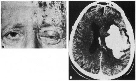
Herpes zoster ophthalmicus behaves like zoster infections elsewhere in the body. The incidence increases with age. Pain, often severe, may precede, be concurrent with, or follow the vesicular skin eruption. Occasionally no skin lesions occur; this is referred to as zoster sine herpete. The vesicular eruption usually involves only one dermatome, but severe systemic eruptions can occur (the latter appear most often in immunocompromised persons or in persons with malignancy). Fever, malaise, headache, and lymphadenopathy may be present at the onset. Fifty percent of cases have ocular involvement, which can be predicted if vesicles appear on the lid margin or if Hutchinson's sign is present.39,40 In 1886, Hutchinson noted that when the nasociliary branch to the tip of the nose is affected, intraocular involvement is likely. Ocular changes include conjunctivitis, episcleritis, scleritis, keratitis, iritis, chorioretinitis, optic neuropathy, glaucoma, ocular motor palsies, and Horner's syndrome.39,41,42 On rare occasions, a contralateral hemiplegia may occur (Fig. 7).39,43–45 A virus-induced angiitis of cerebral vessels has been shown by angiography and pathology in several cases. Although patients with neoplastic or other debilitating diseases are predisposed to the development of herpes zoster infections, a neoplasm is discovered in less than 1% of otherwise healthy persons with cutaneous zoster.

Fig. 7. A: A 70-year-old man with herpes zoster ophthalmicus and a left-cerebral infarction. B: Computed tomography demonstrates a hemorrhagic infarction.

Starting treatment for herpes zoster ophthalmicus should be accomplished as early as possible and ideally within 72 hours. Current recommendations to reduce the incidence of keratitis and possibly postherpetic neuralgia are to use an antiviral drug for 7 to 14 days, lubricating ointment until the cornea is completely healed, but no topical steroids.46 In immunocompetent people, any of the oral antivirals can be used: acyclovir, 800 mg orally five times per day; famciclovir, 500 mg orally three times daily; valacyclovir, 1,000 mg orally three times daily.47–49 Immunocompromised persons should receive intravenous acyclovir.

The gasserian ganglion may also be affected by the spread of infection from a suppurative otitis media. When abducens palsy and pain develop in the face, the symptom complex is called Gradenigo's syndrome (more commonly seen prior to the early use of antibiotics in children with otitis media).50 Lymphoma has also caused this combination of cranial nerve findings.51

Tumors that may involve the ganglion include primary glioma, neurocytoma, neurinoma, malignant schwannoma, meningioma, chordoma, ganglioneuroma, lymphoma, and metastatic tumors. Spread from the nasopharynx, lung, and breast are the most common metastatic tumors and are more frequent than primary tumors.52 Both abnormal sensation and pain can occur in all three of the trigeminal divisions, but motor involvement is unusual. The pain can be constant or paroxysmal, mimicking trigeminal neuralgia. Rarely, loss of the corneal reflex is the only sign. Other cranial nerves may be involved by either enlargement in the middle cranial fossa or growth posteriorly to occupy the cerebellopontine angle. MRI is the most sensitive imaging modality.

TRIGEMINAL ROOT

Tumors involving the root are similar in etiology to those of the gasserian ganglion.53 Pain may be either constant or paroxysmal with all of the features of trigeminal neuralgia. The sensory disturbance usually involves all or most of the face. Often there are other cranial nerve and cerebellar findings.

Tumors or vascular anomalies in the cerebellopontine angle may be large enough to compress the trigeminal root. The most common tumors in this area are vestibular schwannoma, meningioma, and epidermoid cyst.

Blunt head trauma and hydrocephalus are two other conditions that can affect the trigeminal root.54,55

BRAINSTEM

Multiple sclerosis (MS) can produce facial pain, numbness, or other paresthesias. Paroxysmal pain similar in all respects to trigeminal neuralgia occurs in 1% to 2% of patients with MS, which is much higher than the incidence of trigeminal neuralgia in the general population.56 The neuralgia has been found pathologically to be the result of a plaque at the point of entrance of the trigeminal nerve into the brainstem.57 Treatment is discussed in the section on Trigeminal Neuralgia.

Severe constant pain can develop after a lateral medullary infarction (Wallenberg's syndrome) that is the result of a vertebral artery or posterior inferior cerebellar artery occlusion.58 In this syndrome there is usually a dissociated sensory disturbance, with loss of pain and temperature sensation but preservation of light touch and two-point discrimination. Syringobulbia can also produce a similar sensory dissociation.

Intrinsic tumors of the brainstem produce a multitude of neurologic signs, but isolated trigeminal sensory loss virtually never occurs.

Back to Top
THALAMUS, BASAL GANGLIA, AND SENSORY CORTEX
Thalamic lesions can produce either contralateral analgesia or pain, which may be more pronounced in the face than in the rest of the body.

Cranial dystonias can involve the muscles of mastication, producing mouth retraction, jaw opening, or jaw closing. These features are usually part of a more widespread orofacial-cervical dystonia called Meige's syndrome.59,60 The presumed cause of cranial dystonia is an upset in the normal dopamine balance in the basal ganglia and brainstem.61 Treatment is best accomplished with botulinum A toxin injected into the hyperactive muscles.62

Lesions of the inferior postcentral gyrus of the parietal lobe produce numbness in the contralateral side of the face.

SYSTEMIC DISEASES

Trigeminal neuropathy has been reported in numerous systemic diseases, including sarcoidosis, progressive systemic sclerosis, dermatomyositis, Sjögren's syndrome, mixed connective tissue disease, rheumatoid arthritis, and systemic lupus erythematosus.63 Involvement is often bilateral in contrast to trigeminal neuropathy from nonsystemic causes, and sensory deficits with or without pain are invariably noted. When pain is present it is typically constant, but it may be paroxysmal, as in trigeminal neuralgia.

Diabetes mellitus and atherosclerosis cause ocular motor palsies through microvascular ischemia.64 Pain in the eye and forehead, which is likely the result of a similar microvascular process involving the trigeminal nerve, may also occur and may be severe in nature.65 Although sensation is usually normal, the authors have examined one diabetic patient who developed a pupil-sparing oculomotor nerve palsy associated with pain and a sensory deficit in the ophthalmic division. Computed tomography was normal. The sensory deficit and oculomotor palsy completely resolved over a 6-week period.

Back to Top
FACIAL PAIN
Several of the facial pain syndromes include trigeminal neuralgia, cluster

headache, temporomandibular joint disease, postherpetic neuralgia, and atypical (chronic) facial pain. Although a clear distinction may not always be possible, it is useful to classify facial pain as either constant or paroxysmal. Constant pain is frequently symptomatic of an underlying structural lesion. Often there are paroxysms of more severe pain on top of the constant pain, but pure trigeminal neuralgia should be pain free between the painful shocks, and there should be no sensory or motor deficit.

TRIGEMINAL NEURALGIA (TIC DOULOUREUX)

Trigeminal neuralgia is characterized by paroxysms of severe lancinating pain in the distribution of the trigeminal nerve. The maxillary and mandibular divisions are more commonly involved than the ophthalmic, but all three divisions or any combination of them can be affected. Bilateral involvement occurs in 3% to 12% of cases, depending on the series.66 Women are affected more commonly than men, and the average age of onset is around 50 years. The pain occurs more often on the right in most but not all series.67,68 Familial occurrence is rare.69

The pain occurs without prodromata and typically lasts only seconds, although aftershocks may occur for several minutes. The pain is often described as an electric shock sensation. The interval between shocks can be just a few seconds or as long as a few hours. After several days to weeks, the pain remits, and it may be weeks or years before another episode. Between episodes the patient is free of pain. Triggers include touch, speech, chewing, and even the wind. The avoidance of these triggers can be so extreme that dehydration and malnutrition may occur.

Diagnosis of trigeminal neuralgia is based on the history. Examination during an attack may demonstrate hypalgesia or hyperesthesia in the affected area, but no abnormalities should be detected when the patient is pain free. The different causes of trigeminal neuralgia can be sorted out with the use of appropriate imaging techniques. Magnetic resonance imaging (MRI) including angiographic sequences has become the premier modality to separate microvascular compression from tumor, arteriovenous malformation, and MS.70 Occasionally no cause is identified. When the cause is MS, patients are usually younger, more often have bilateral disease, and have other evidence of demyelination by history and examination. Approximately 2% to 4% of trigeminal neuralgia cases are caused by MS.56 Tumor types are mainly trigeminal neurinoma, vestibular schwannoma, meningioma, and cholesteatoma.71 Evidence for dysfunction of neighboring cranial nerve VII or VIII may suggest a tumor.

Microvascular compression as a cause of trigeminal neuralgia was first postulated by Dandy in 1934.72 Estimates as to its frequency have ranged between 11% and 96%, with most recent operative series reporting approximately 90%.67,70,73,74 The offending vessel is most often the superior cerebellar artery, although many other arteries and, less frequently, veins have been reported.75 The pathophysiology is thought to be either pulsating trauma or cross-compression of the vessel on the nerve, resulting in demyelination and spontaneous nerve firing., Both peripheral and central mechanisms of pain generation have also been implicated. Vessels are found at the root entry zone far more often than at other locations along the nerve. The theory of microvascular compression is not accepted by all. Several arguments are laid out by Adams,76 including an alternative reason for pain relief after surgery.

In the absence of a tumor, medical management of trigeminal neuralgia should always precede the consideration of surgical intervention, because spontaneous remission is high. The common thread among drugs now in use is their ability to depress spontaneous excitatory transmission along the trigeminal pathway. Carbamazepine (Tegretol) is the initial drug of choice and is effective usually within 1 to 2 days in 70% to 90% of cases.77,78 Twenty-five percent of patients who initially respond will become refractory to the drug, and an additional 5% to 19% will develop side effects that necessitate its withdrawal. In our experience, oxcarbazepine is an effective cousin to carbamazepine typically with fewer side effects.79 Phenytoin is an effective, inexpensive drug.78,80 Other medications that have been used with some success include Baclofen, clonazepam, divalproex sodium, and lamotrigine.78,81 A small series of MS patients had some benefit with misoprostol.82 Finally, nonspecific analgesics and anti-inflammatories such as nonsteroidal antiinflammatory drugs, prednisone, and opiates have their use in acute management. Only carbamazepine is approved by the Food and Drug Administration (FDA).

The literature suggests that 25% to 50% of patients with trigeminal neuralgia will eventually need surgery because of either poor pain control or medication intolerance.67 Several different surgical procedures are available.83 The two most common and effective procedures are microvascular decompression and percutaneous radiofrequency thermocoagulation of the ganglion. Other surgical options that will not be discussed further include sectioning of the trigeminal root, trigeminal tractotomy near the cervicomedullary junction, retrogasserian glycerol rhizotomy, balloon compression of the ganglion, and peripheral cryotherapy.

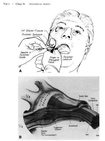
Selective percutaneous thermocoagulation of the gasserian ganglion (gangliolysis) can be performed as an outpatient procedure or with overnight hospitalization.84 An electrode, viewed radiographically, is placed through the foramen ovale into the gasserian ganglion, and its position is adjusted according to the patient's response to gentle electrical stimulation (Fig. 8). Graded radiofrequency current is then applied to produce differential destruction of pain fibers while sparing light touch fibers. Short-acting barbiturates are given to the patient for the painful parts of the procedure but otherwise it is done with the patient awake. Immediate relief of pain is achieved in more than 90%.68,84,85 In a prospective study by Taha and colleagues,68 the recurrence rate was 15% at 5 years, 22% at 10 years, and 25% at 14 years. Repeat gangliolysis, if needed, was moderately successful. Anesthesia of the face and cornea, dysesthesias, and trigeminal motor weakness are the main drawbacks to gangliolysis. In fact, there is a direct relationship between the degree of facial sensory loss and the long-term success of the procedure.68 The sensory changes are annoying or painful in 5% to 25% of patients, and this, too, is directly related to the degree of sensory loss. Therefore, the greatest success is achieved through the procedure's greatest drawback. Motor weakness affects around 20% of patients and in most is transient.86 This procedure is particularly suited for patients opposed to craniotomy, patients with MS, and elderly or medically fragile patients who are not good surgical candidates for microvascular decompression.

Fig. 8. Technique of percutaneous controlled thermocoagulation of the trigeminal ganglion. A: Needle placement. B: Position of needle in the ganglion. (From Tew JM, Keller JT: Percutaneous rhizotomy in the treatment of intractable facial pain. In Lee JF (ed): Pain Management—Symposium on the Neurosurgical Treatment of Pain. Baltimore: Williams & Wilkins, 1977:150–155.)

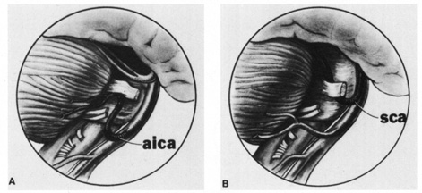
Microvascular decompression surgery is carried out by performing a suboccipital craniotomy. The operating microscope is used to identify anomalous blood vessels that may be compressing the trigeminal root, and a nonabsorbable sponge is placed over the nerve for its protection (Fig. 9). In a large series of 1,185 patients, immediate complete relief of pain was reported in 82%, whereas another 16% had partial relief.67 Long-term benefits for complete relief were noted in 75% at 1 year and 64% at 10 years. The percentage of patients with recurrence after complete relief is similar to that of patients undergoing radiofrequency gangliolysis if one accounts for the smaller number of patients getting immediate relief from decompression. In the same series,67 complications were transient in 25% and permanent in 4%, including two deaths. The main advantages to microvascular decompression are that it is directed at what may be the actual cause of trigeminal neuralgia, and there is much less chance of permanent facial numbness.

Fig. 9. Vascular compression of the trigeminal root by (A) the anterior inferior cerebellar artery (aica) and (B) the superior cerebellar artery (sca). (From Jannetta PJ, Zorub DS: Microvascular decompression for trigeminal neuralgia. In Buchheit WA, Truex RC (eds): Surgery of the Posterior Fossa. New York: Raven Press, 1979:146–147.)

Stereotactic radiosurgery (gamma knife, linear accelerator [LINAC]) is the newest addition and least invasive treatment method for trigeminal neuralgia. This treatment, like gangliolysis, can be used for patients with symptomatic diseases such as MS and trauma, in contrast to microvascular decompression. Radiation is directed at the root entry zone of the trigeminal nerve. Several studies of medically and surgically refractory patients with a variety of causes have been done. Similar results were found with patients having no pain or a good response in 77% to 88%.88,89 Recurrence of pain, and facial numbness occurred between 10% and 25%.87,88

Only a randomized study involving patients undergoing a first surgical procedure will determine the best treatment if medications fail.

Back to Top
CLUSTER HEADACHE
Cluster headache is characterized by the acute onset of severe, unilateral, constant pain that is centered around the eye in either the orbit, forehead, temple, or cheek90 and may spread to the ipsilateral head or neck. The pain causes patients to become restless and very active. The name comes from the clustering of attacks, usually one or two times per year lasting weeks to several months. In between attacks there are pain-free intervals that can span many years. Some patients have chronic cluster headache in which no remissions occur. Autonomic signs or symptoms ipsilateral to the pain are required for the diagnosis of cluster headache. At least one of the following is needed: conjunctival injection, lacrimation, nasal congestion, rhinorrhea, forehead or facial sweating, miosis, ptosis, or eyelid edema. Signs of Horner's syndrome (oculosympathetic paresis), with accompanying miosis and ptosis, require differentiation from carotid artery dissection and Raeder's paratrigeminal neuralgia (see later).

Another interesting feature of the pain in cluster headache is the chronobiology. Attacks usually occur at the same time of the day or night anywhere from one to eight times in a 24-hour period. This, and positron emission tomography (PET) scan data, has led to the idea that the hypothalamus is important in the pathogenesis along with the trigeminovascular system.91 Each attack lasts 15 minutes to several hours and resolves as quickly as it comes on. Alcohol is often reported as a trigger. Head injury has been reported to precipitate cluster headache.92 Men are much more likely than women to have cluster headaches with onset in the third or fourth decade, but onset at any age is possible.

Treatment can be separated into medications used in the acute setting and those used for prophylaxis.93,94 Options for acute therapy include oxygen, fast acting triptans, dihydroergotamine, ergotamine, and fast acting opiates. Only subcutaneous sumatriptan is FDA approved. Prophylactic medications include prednisone, lithium, verapamil, methysergide, sodium valproate, gabapentin, and topiramate. Future medications may include melatonin and botulinum toxin. Finally, in rare cases resistant to standard therapy, consideration should be given to selective percutaneous thermocoagulation of the trigeminal ganglion, and more recently, hypothalamic deep brain stimulation.95

Cluster headaches can be contrasted with migraine headaches. The latter are more common in women, do not cluster, rarely have autonomic symptoms, cause people to seek a quiet room in which to lie down, have a throbbing quality, and do not respond to oxygen.

Back to Top
RAEDER'S PARATRIGEMINAL NEURALGIA
When severe facial or head pain is associated with ipsilateral ptosis and miosis secondary to sympathetic denervation, the disorder is called Raeder's paratrigeminal neuralgia.38,96 Localization in all cases is thought to involve the third-order sympathetic neurons along the internal carotid artery or in the cavernous sinus. The syndrome is categorized as type 1 when parasellar cranial nerve palsies (III, IV, V, and VI) are also present, and as type 2 when just pain and oculosympathetic paresis occur. Many authors have stated that the complete Horner's syndrome, which includes anhidrosis, is not found in Raeder's syndrome. This is only partly true. Most of the sympathetic fibers innervating sweat glands travel with the external rather than the internal carotid. Sweat glands of the medial forehead, however, are innervated by internal carotid fibers; therefore, anhidrosis of this area is not uncommon.97

The etiology of type 1 Raeder's paratrigeminal neuralgia is varied. Most cases are caused by middle cranial fossa masses, but neurosyphilis, aneurysm, and trauma have been implicated.98,99 A demyelinating, inflammatory, or infectious pathophysiology is blamed for type 2 cases. It is very important to consider and rule out carotid artery dissection early because morbidity from cerebral ischemia may be avoidable.

Cluster headache may manifest an oculosympathetic paresis, as noted earlier, and could be confused with type 2 Raeder's syndrome. There are differences, however, that will help separate the two disorders.96,97 Cluster headache is characterized by nausea and vomiting, photophobia, other autonomic symptoms, recurrence, precipitation by alcohol, severe pain along with breaks in which the patient is pain free, and response to oxygen, steroids, and other medications. Differentiation must also be made from the Tolosa-Hunt syndrome.

Back to Top
TEMPOROMANDIBULAR JOINT DISEASE
Temporomandibular joint misalignment, which is usually secondary to malocclusion, can produce facial pain that radiates to the forehead, vertex, or occiput.100,101 The pain may be intermittent or constant; it frequently worsens as the day progresses, and it is exacerbated by chewing. Laxity in the mandibular articulation may be palpable. Relief can be provided by physical therapy, mouth orthotics, anti-inflammatory drugs, muscle relaxants, chronic pain medications such as nortriptyline, and surgery.101
Back to Top
POSTHERPETIC NEURALGIA
Herpes zoster infection, or shingles, is the result of the reactivation of latent varicella zoster virus that has remained quiet in the sensory ganglia of the body since a primary chickenpox infection. The rash usually occupies one or two dermatomes but rarely disseminates; it remains unilateral and is most often thoracic. The ophthalmic division of the trigeminal nerve is the most common cranial nerve site, and the percentage of cases with ophthalmic involvement increases with age. In addition to age, immunosuppression is a major risk factor. Of the potential complications of herpes zoster, postherpetic neuralgia is the most common.

One definition of postherpetic neuralgia is pain that persists 1 month or more after the skin lesions have healed. This occurs overall in approximately 10% of patients with age as the biggest risk factor.102 Trigeminal involvement is associated with a higher incidence of postherpetic neuralgia. The pain is described as a constant, intense, burning sensation, but it may be episodic, lasting about 30 minutes at a time. Hypesthesia or hyperalgesia may be detected in the involved dermatome. Fortunately, most people with postherpetic neuralgia have resolution of their pain within 1 year.

Pain relief has been reported with gabapentin, tricyclic antidepressants, topical lidocaine, capsaicin, , carbamazepine, phenytoin, valproic acid, nonsteroidal anti-inflammatory drugs, narcotics, counterirritation, biofeedback, and transcutaneous electrical nerve stimulation.49,103,104 First-line therapy is usually gabapentin (300 to 3600 mg divided per day) based on a large randomized study.105 In refractory cases, surgical procedures to interrupt or modify central pain pathways have been successful.49,106 Kost and Straus,49 in a review of the subject, provide a flow chart to help with management.

Efforts at prevention of postherpetic neuralgia are as important as the trials evaluating medications for pain relief. Although the results of early trials using steroids during the acute vesicular rash were encouraging, subsequent trials have failed to show any benefit.107 However, steroids are beneficial in shortening the duration of acute pain that accompanies every eruption and in improving quality of life; therefore they can be recommended as treatment if given within the first 72 hours for persons older than 50 years of age with no contraindications.108 A typical dose is 40 to 60 mg of prednisone tapered over 3 weeks. Antiviral agents have been studied, and although not all trials came to the same conclusion, there appears to be a reduction in acute pain and postherpetic neuralgia.49,109,110 Seven days of acyclovir, famciclovir, or valacyclovir is recommended if started early.

Back to Top
ATYPICAL FACIAL PAIN
Some cases of facial pain are not explained by any of the diseases usually responsible, even after a careful search for a structural lesion with MRI. When this occurs, the authors resort to the diagnosis of atypical facial pain.111 Many articles correctly comment that “atypical” does not describe what the problem is. Chronic facial pain is preferred by some and is one category under the larger heading of orofacial pain syndrome which also includes temporomandibular joint dysfunction, odontalgia, and burning-mouth syndrome.112 Etiologies to be considered and ruled out include the following: trigeminal neuralgia; cluster headache; temporomandibular joint disease; postherpetic neuralgia; Tolosa-Hunt syndrome; MS; disorders of the teeth, sinuses, or cervical spine; tumors or other compressive lesions of the trigeminal nerve; carotid dissection; carotidynia; and giant cell arteritis.111,113

The pain is usually constant, deep, and felt as a burning or aching. It is not limited to a single dermatome and may spread across the midline. In addition to the constant pain, there may be provocable, lightening-like pains of short duration, making a clear separation from trigeminal neuralgia difficult.

The several theories concerning pathogenesis incorporate the observations that most of the patients who suffer from atypical (chronic) facial pain are middle-aged women and that a recent history of dental or sinus surgery is common. Pain may result from local nerve trauma at the time of surgery, psychosomatic disease, trigeminovascular alterations as with migraine, and complex regional pain syndrome.111,114

This condition is often resistant to treatment. A combination of psychotherapy, antidepressants, anticonvulsants, and benzodiazepines may be of value. From a larger study using stereotactic radiosurgery for facial pain, the subset of patients with atypical facial pain had no improvement.88

Back to Top
OCULAR PAIN
Pain in and around the eye may be caused by ocular disease itself, or it may be referred from another structure. Corneal foreign bodies, epithelial defects or inflammation, iridocyclitis, acute angle-closure glaucoma, optic neuritis, myositis, and orbital inflammations are some examples.115

Any disorder of the trigeminal nerves, ganglion, or root can produce pain that is felt primarily in or around the eye. The description of the pain and the detection of a sensory deficit or other neurologic abnormalities will provide clues to the diagnosis.

Meningeal irritation may produce pain that is referred to the eye because branches of the ophthalmic division supply the dura mater. This is the probable cause of the pain associated with oculomotor nerve palsy caused by an aneurysm and the pain associated with homonymous hemianopia resulting from posterior cerebral artery occlusion.116

Often the ophthalmologist is presented with a patient who complains of pain in or about the eye and in whom there is no apparent ocular or neurologic cause after a full examination. Sjögren's syndrome should be considered if there is a complaint of a dry eye or mouth. Sinus films should be taken but are rarely positive without a suggestive sinus history from the patient. Occasionally the description of the pain is typical of tension, migraine, or cluster headaches. Referred pain can come from cervical disease or posterior fossa masses.117,118 Giant cell arteritis is often considered as a cause of pain around the eye, but it would be extremely unusual in the absence of other symptoms or signs of arteritic disease, such as poor appetite, weight loss, malaise, polymyalgia rheumatica, jaw claudication, ischemic optic neuropathy, and elevated sedimentation rate.

Back to Top
THE PAINFUL BLIND EYE
Chronic ocular pain may occur in blind, severely disturbed eyes with end-stage glaucoma, long-standing retinal detachment, chronic hypoxia, severe infection or inflammation, or previous trauma. Treatment of the pain is often difficult. Topical cycloplegic medications may provide some relief. Radiofrequency percutaneous thermocoagulation of the gasserian ganglion (by the same method described for trigeminal neuralgia) may have long-term effects.119 When other methods for relief of pain in the blind eye fail, enucleation should be considered.
Back to Top
ACKNOWLEDGMENT
The authors thank Dr. Roy Beck for his contribution to this chapter.
Back to Top
REFERENCES

1. Burton H: Somatosensory sensations from the eye. In Moses RA (ed.): Adler's Physiology of the Eye. St. Louis: CV Mosby, 1981:63–68

2. Ray BS, Wolff HG: Experimental studies on headache. Pain-sensitive structures of the head and their significance in headache. Arch Surg 41:813, 1940

3. Feindel W, Penfield W, McNaughton F: The tentorial nerves and localization of intracranial pain in man. Neurology 10:555, 1960

4. Wirth FP, Van Buren JM: Referral of pain from dural stimulation in man. J Neurosurg 34:630, 1971

5. Moonie GT, Rees DL, Elton D: The oculocardiac reflex during strabismus surgery. Can Anaesth Soc J 11:621, 1964

6. Lang S, Lanigan DT, van der Wal M: Trigeminocardiac reflexes: maxillary and mandibular variants of the oculocardiac reflex. Can J Anaesth 38:757, 1991

7. Barnard NA, Bainton R: Bradycardia and the trigeminal nerve. J Craniomaxillofac Surg 18:359, 1990

8. Haerer AF: DeJong's The Neurologic Examination, 5th ed. Philadelphia: JB Lippincott, 1992:165–179

9. Kimura J: The blink reflex. In Kimura J: Electrodiagnosis in Diseases of Nerve and Muscle: Principles and Practice, 2nd ed. Philadelphia: FA Davis, 1989:307–331

10. Ongerboer de Visser BW, Melchelse K, Megens PHA: Corneal reflex latency in trigeminal nerve lesions. Neurology 27:1164, 1977

11. Kimura J: Somatosensory and motor evoked potentials. In Kimura J: Electrodiagnosis in Diseases of Nerve and Muscle: Principles and Practice, 2nd ed. Philadelphia: FA Davis, 1989:395–397

12. Kimura J: H, T, masseter and other reflexes. In Kimura J: Electrodiagnosis in Diseases of Nerve and Muscle: Principles and Practice, 2nd ed. Philadelphia: FA Davis, 1989:361–363

13. Goor C, Ongerboer de Visser BW: Jaw and blink reflexes in trigeminal nerve lesions: an electrodiagnostic study. Neurology 26:95, 1976

14. Taverner D: The localization of isolated cranial nerve lesions. In Vinken PJ, Bruyn GW (eds.): Handbook of Clinical Neurology, Vol. 2. Amsterdam: North-Holland, 1969:54–59

15. Martin XY, Safran AB: Corneal hypoesthesia. Surv Ophthalmol 33:28, 1988

16. De Haas EBH: Desiccation of cornea and conjunctiva after sensory denervation. Arch Ophthalmol 67:439, 1962

17. Mackie IA: Role of the corneal nerves in destructive diseases of the cornea. Trans Ophthalmol Soc UK 98:343, 1978

18. Duke-Elder S, Leigh AG: Diseases of the outer eye. In Duke-Elder S (ed.): System of Ophthalmology, Vol. 8. St. Louis: CV Mosby, 1965:809

19. Rowbotham GF: Acute Injuries of the Head. Baltimore: Williams & Wilkins, 1964:422–424

20. Goldstein NP, Gibilisco JA, Rushton JG: Trigeminal neuropathy and neuritis: a study of etiology with emphasis on dental causes. JAMA 184:458, 1963

21. Ferdousi AM, MacGregor AJ: The response of the peripheral branches of the trigeminal nerve to trauma. Int J Oral Surg 14:41, 1985

22. Rose GE, Wright JE: Trigeminal sensory loss in orbital disease. Br J Ophthalmol 78:427, 1994

23. Selby G: Diseases of the fifth cranial nerve. In Dyck PJ, Thomas PK, Lambert EH, Bunge R (eds.): Peripheral Neuropathy, 2nd ed., Vol. II. Philadelphia: WB Saunders, 1984:1224–1265

24. Blau JN, Harris M, Kennett S: Trigeminal sensory neuropathy. N Engl J Med 281:873, 1969

25. Peñarrocha M, Alfaro A, Bagán JV, et al: Idiopathic trigeminal sensory neuropathy. J Oral Maxillofac Surg 50:472, 1992

26. Horowitz SH: Isolated facial numbness: clinical significance and relation to trigeminal neuropathy. Ann Intern Med 80:49, 1974

27. Spillane JD, Wells CEC: Isolated trigeminal neuropathy: a report of 16 cases. Brain 82:391, 1959

28. Hutchins LG, Harnsberger HR, Hardin CW, et al: The radiologic assessment of trigeminal neuropathy. AJR Am J Roentgenol 153:1275, 1989

29. Rorick MB, Chandar K, Colombi BJ: Inflammatory trigeminal sensory neuropathy mimicking trigeminal neurinoma. Neurology 46:1455, 1996

30. Cruccu G, Inghilleri M, Berardelli A, et al: Pathophysiology of hemimasticatory spasm. J Neurol Neurosurg Psychiatry 57:43, 1994

31. Thompson PD, Carroll WM: Hemimasticatory spasm—a peripheral paroxysmal cranial neuropathy? J Neurol Neurosurg Psychiatry 46:274, 1983

32. Trobe JD, Glaser JS, Post JD: Meningiomas and aneurysms of the cavernous sinus: neuro-ophthalmologic features. Arch Ophthalmol 96:457, 1978

33. Inoue T, Fukui M, Matsushima T, et al: Neurinoma in the cavernous sinus: report of two cases. Neurosurgery 27:986, 1990

34. Tolosa E: Periarteritic lesions of the carotid siphon with the clinical features of a carotid infraclinoidal aneurysm. J Neurol Neurosurg Psychiatry 17:300, 1954

35. Hunt WE, Meagher JN, LeFever HE, et al: Painful ophthalmoplegia: its relation to indolent inflammation of the cavernous sinus. Neurology 11:56, 1961

36. Juncos JL, Beal MF: Idiopathic cranial polyneuropathy: a fifteen-year experience. Brain 110:197, 1987

37. Spector RH, Schwartzman RJ: Benign trigeminal and facial neuropathy. Arch Intern Med 135:992, 1975

38. Raeder JG: Paratrigeminal paralysis of oculo-pupillary sympathetics. Brain 47:149, 1924

39. Marsh RJ, Cooper M: Ophthalmic herpes zoster. Eye 7:350, 1993

40. Tomkinson A, Roblin DG, Brown MJKM: Hutchinson's sign and its importance in rhinology. Rhinology 33:180, 1995

41. Roberts TV, Francis IC, Kappagoda MB, et al: Herpes zoster chorioretinopathy. Eye 9:594, 1995

42. Smith EF, Santamarina L, Wolintz AH: Herpes zoster ophthalmicus as a cause of Horner syndrome. J Clin Neuroophthalmol 13:250, 1993

43. Acers TE: Herpes zoster ophthalmicus with contralateral hemiplegia. Arch Ophthalmol 71:371, 1964

44. Kuroiwa Y, Furukawa T: Hemispheric infarction after herpes zoster ophthalmicus: Computed tomography and angiography. Neurology 31:1030, 1981

45. NcNeil JD, Horowitz M: Contralateral hemiplegia complicating herpes zoster ophthalmicus. J R Soc Med 84:501, 1991

46. Severson EA, Baratz KH, Hodge DO, et al: Herpes zoster ophthalmicus in Olmsted County, Minnesota: have systemic antivirals made a difference. Arch Ophthalmol 121:386, 2003

47. Tyring S, Engst R, Corriveau C, et al: Famciclovir for ophthalmic zoster: a randomized aciclovir controlled study. Br J Ophthalmol. 85:576, 2001

48. Colin J, Prisant O, Cochener B, et al: Comparison of the efficacy and safety of valaciclovir and acyclovir for the treatment of herpes zoster ophthalmicus. Ophthalmology 107:1507, 2000

49. Kost RG, Straus SE: Postherpetic neuralgia—pathogenesis, treatment, and prevention. N Engl J Med 335:32, 1996

50. Gradenigo G: A special syndrome of endocranial otitic complications (paralysis of the motor oculi externus of otitic origin). Ann Otol 13:637, 1904

51. Tutunuoglu S, Uran N, Kavas I, et al: Gradenigo syndrome: a case report. Pediatr Radiol 23:556, 1993

52. Hedeman LS, Lewinsky BS, Lochridge GK, et al: Primary malignant schwannoma of the gasserian ganglion: report of two cases. J Neurosurg 48:279, 1978

53. Tancioni F, Gaetani P, Villani L, et al: Neurinoma of the trigeminal root and atypical trigeminal neuralgia: case report and review of the literature. Surg Neurol 44:36, 1995

54. Summers CG, Wirtschafter JD: Bilateral trigeminal and abducens neuropathies following low-velocity, crushing head injury. J Neurosurg 50:508, 1979

55. Maurice-Williams RS, Pilling J: Trigeminai sensory symptoms associated with hydrocephalus. J Neurol Neurosurg Psychiatry 40:641, 1977

56. Hooge JP, Redekop WK: Trigeminal neuralgia in multiple sclerosis. Neurology 45:1294, 1995

57. Olafson RA, Rushton JG, Sayre GP: Trigeminal neuralgia in a patient with multiple sclerosis: an autopsy report. J Neurosurg 24:755, 1966

58. Currier RD, Giles CL, DeJong RN: Some comments on Wallenberg's lateral medullary syndrome. Neurology 11:778, 1961

59. Meige H: Les convulsions de la face, une forme clinique de convulsion faciale, bilaterale et mediane. Rev Neurol (Paris) 20:437, 1910

60. Marsden CD: Blepharospasm-oromandibular dystonia syndrome (Brueghel's syndrome): a variant of adult onset torsion dystonia? J Neurol Neurosurg Psychiatry 39:1204, 1976

61. Berardelli A, Rothwell JC, Marsden CD: Pathophysiology of blepharospasm and oromandibular dystonia. Brain 108:593, 1985

62. Jankovic J, Orman J: Botulinum A toxin for cranial-cervical dystonia: a double blind, placebo-controlled study. Neurology 37:616, 1987

63. Olney RK: AAEM minimonograph No. 38: neuropathies in connective tissue disease. Muscle Nerve 15:531, 1992

64. Walsh FB, Hoyt WF: Clinical Neuro-ophthalmology. Baltimore: Williams & Wilkins, 1969:350–433, 1351–1361

65. Ross AT: Recurrent cranial nerve palsies in diabetes mellitus. Neurology 12:180, 1962

66. Brisman R: Bilateral trigeminal neuralgia. J Neurosurg 67:44, 1987

67. Barker FG, Jannetta PJ, Bissonette DJ, et al: The long-term outcome of microvascular decompression for trigeminal neuralgia. N Engl J Med 334:1077, 1996

68. Taha JM, Tew JM, Buncher CR: A prospective 15-year follow up of 154 consecutive patients with trigeminal neuralgia treated by percutaneous stereotactic radiofrequency thermal rhizotomy. J Neurosurg 83:989, 1995

69. Herzberg L: Familial trigeminal neuralgia. Arch Neurol 37:285, 1980

70. Meaney JFM, Eldridge PR, Dunn LT, et al: Demonstration of neurovascular compression in trigeminal neuralgia with magnetic resonance imaging. J Neurosurg 83:799, 1995

71. Nomura T, Ikezaki K, Matsushima T, et al: Trigeminal neuralgia: differentiation between intracranial mass lesions and ordinary vascular compression as causative lesions. Neurosurg Rev 17:51, 1994

72. Dandy WE: Concerning the cause of trigeminal neuralgia. Am J Surg 24:447, 1934

73. Adams CBT, Kaye AH, Teddy PJ: The treatment of trigeminal neuralgia by posterior fossa microsurgery. J Neurol Neurosurg Psychiatry 45:1020, 1982

74. Hamlyn PJ, King TT: Neurovascular compression in trigeminal neuralgia: a clinical and anatomical study. J Neurosurg 76:948, 1992

75. Jannetta PJ: Microvascular decompression of the trigeminal nerve root entry zone. In Rovit RL, Murali R, Jannetta PJ (eds.): Trigeminal Neuralgia. Baltimore: Williams & Wilkins, 1990:201–222

76. Adams CBT: Microvascular compression: an alternative view and hypothesis. J Neurosurg 57:1, 1989

77. Blom S: Tic douloureux treated with new anticonvulsant. Arch Neurol 9:285, 1963

78. Green MW, Selman JE: Review article: the medical management of trigeminal neuralgia. Headache 31:588, 1991

79. Zakrzewska JM, Patsalos PN: Long-term cohort study comparing medical (oxcarbazepine) and surgical management of intractable trigeminal neuralgia. Pain 95:259, 2002

80. Iannone A, Baker AB, Morrell F: Dilantin in the treatment of trigeminal neuralgia. Neurology 8:126, 1958

81. Zakrzewska JM, Chaudry Z, Numikko TJ, et al: Lamotrigine (Lamictal) in refractory trigeminal neuralgia: results from a double-blind placebo controlled crossover trial. Pain 73:223, 1997

82. Reder AT, Arnason BGW: Trigeminal neuralgia in multiple sclerosis relieved by a prostaglandin E analogue. Neurology 45:1097, 1995

83. Loeser JD: What to do about tic douloureux. JAMA 239:1153, 1978

84. Sweet WH, Wepsic JG: Controlled thermocoagulation of trigeminal ganglion and rootlets for differential destruction of pain fibers. Part 1: Trigeminal neuralgia. J Neurosurg 39:143, 1974

85. Apfelbaum RI: A comparison of percutaneous radiofrequency trigeminal neurolysis and microvascular decompression of the trigeminal nerve for the treatment of tic douloureux. Neurosurgery 1:16, 1977

86. Rovit RL: Percutaneous radiofrequency thermal coagulation of the gasserian ganglion. In Rovit RL, Murali R, Jannetta PJ (eds.): Trigeminal Neuralgia. Baltimore: Williams & Wilkins, 1990:109–136

87. Kondziolka D, Perez B, Flickinger JC, et al: Gamma knife radiosurgery for trigeminal neuralgia: results and expectations. Arch Neurol 55:1524, 1998

88. Smith ZA, DeSalles AAF, Frighetto L, et al: Dedicated linear accelerator radiosurgery for the treatment of trigeminal neuralgia. J Neurosurg 99:511, 2003

89. Young RF, Vermeulen SS, Grimm P, et al: Gamma knife radiosurgery for treatment of trigeminal neuralgia: idiopathic and tumor related. Neurology 48:608, 1997

90. Horton BT, MacLean AR, Craig WM: A new syndrome of vascular headache. Results of treatment with histamine: Preliminary report. Proceedings of the Staff Meetings of the Mayo Clinic 14:257, 1939

91. Kudrow L: The pathogenesis of cluster headache. Curr Opin Neurol 7:278, 1994

92. Turkewitz LJ, Wirth O, Dawson GA, et al: Cluster headache following head injury: a case report and review of the literature. Headache 32:504, 1992

93. Matharu MS, Boes CJ, Goadsby PJ: Management of trigeminal autonomic cephalgias and hemicrania continua. Drugs 63:May A, Leone M: Update on cluster headache. Curr Opin Neurol 16:333, 2003

94. 1637, 2003

95. Leone M, Franzini A, Broggi G, et al: Hypothalamic deep brain stimulation for intractable chronic cluster headache: a 3-year follow-up. Neurol Sci 24:S143, 2003

96. Grimson BS, Thompson HS: Raeder's syndrome. A clinical review. Surv Ophthalmol 24:199, 1980

97. Sjaastad O, Elsås T, Shen J, et al: Raeder's syndrome: “anhidrosis,” headache, and a proposal for a new classification. Funct Neurol 9:215, 1994

98. Kashihara K, Ito H, Yamamoto S, et al: Raeder's syndrome associated with intracranial internal carotid artery aneurysm. Neurosurgery 20:49, 1987

99. Watanabe K, Tanahashi N, Nara M: Neurosyphilis presenting with Raeder's syndrome. Ann Neurol 25:418, 1989

100. Costen JB: Neuralgias and ear symptoms associated with disturbed function of the temporomandibular joint. JAMA 107:252, 1936

101. Kirk WS: Chronic temporomandibular joint disease and head pain: response to surgery. N C Med J 54:30, 1993

102. Helgason S, Petursson G, Gudmundsson S, et al: Prevalence of postherpetic neuralgia after a first episode of herpes zoster: prospective study with long term follow up. BMJ 321:794, 2000

103. Rowbotham MC, Davies PS, Fields HL: Topical lidocaine gel relieves postherpetic neuralgia. Ann Neurol 37:246, 1995

104. Dubinsky RM, Kabbani H, El-Chami Z, et al: Practice parameter: treatment of postherpetic neuralgia. An evidenced-based report of the quality standards subcommittee of the American Academy of Neurology. Neurology 63:959, 2004

105. Rowbotham M, Harden N, Stacey B, et al: Gabapentin for the treatment of postherpetic neuralgia: a randomized controlled trial. JAMA 280:1837, 1998

106. Loeser JD: Herpes zoster and postherpetic neuralgia. Pain 25:149, 1986

107. Keczkes K, Basheer AM: Do corticosteroids prevent post-herpetic neuralgia? Br J Dermatol 102:551, 1980

108. Whitley RJ, Weiss H, Gnann JW Jr, et al: Acyclovir with and without prednisone for the treatment of herpes zoster. A randomized, placebo-controlled trial. The National Institute of Allergy and Infectious Diseases Collaborative Antiviral Study Group. Ann Intern Med 125:376, 1996

109. Wood MJ, Johnson RW, McKendrick MW, et al: A randomized trial of acyclovir for 7 days or 21 days with and without prednisolone for treatment of acute herpes zoster. N Engl J Med 330:896, 1994

110. Jackson JL, Gibbons R, Meyer G, et al: The effect of treating herpes zoster with oral acyclovir in preventing postherpetic neuralgia. A meta-analysis. Arch Intern Med 157:909, 1997

111. Türp JC, Gobetti JP: Trigeminal neuralgia versus atypical facial pain: a review of the literature and case report. Oral Surg Oral Med Oral Pathol Oral Radiol Endod 81:424, 1996

112. Madland G, Feinmann C: Chronic facial pain: a multidisciplinary problem. J Neurol Neurosurg Psychiatry 71:716, 2001

113. Cusick JF: Atypical trigeminal neuralgia. JAMA 245:2328, 1981

114. Weddington WW, Blazer D: Atypical facial pain and trigeminal neuralgia: a comparison study. Psychosomatics 20:348, 1979

115. Rosenblatt MA, Sakol PJ: Ocular and periocular pain. Otolaryngol Clin North Am 22:1173, 1989

116. Knox DL, Cogan DG: Eye pain and homonymous hemianopia. Am J Ophthalmol 54:1091, 1962

117. Kerr FWL: A mechanism to account for frontal headache in cases of posterior fossa tumors. J Neurosurg 18:605, 1961

118. Edmeads J: Headaches and head pains associated with diseases of the cervical spine. Med Clin North Am 62:533, 1978

119. Rosenberg M, Hoyt CS, King JS, et al: Treatment of chronic ocular pain by selective thermocoagulation of the trigeminal ganglion. Am J Ophthalmol 91:526, 1981

Back to Top