|
|
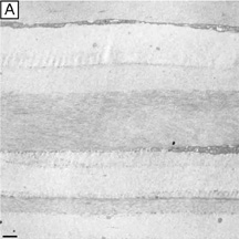
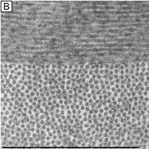
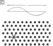
| Fig. 10. (A) Low magnification (4,750×) TEM of predominantly orthogonally stacked lamellae in the middle third of cellular corneal stroma. (B) Higher magnification (72,500×) TEM of two lamellae in the middle third of cellular corneal stroma. One lamellae is in longitudinal view (top) and other is in cross-sectional view (bottom). Notice the uniformly 25-nm diameter collagen fibrils and the 40-nm diameter interfibrillar spaces that demonstrate only a short-range order (i.e., slight variability), but do not form a true crystalline lattice. (C) Cross-sectional diagram of collagen fibrils arranged in a true crystalline lattice arrangement. Size of a wavelength of light is shown above for comparison. Bars = 1 μm. (From Maurice DM. The structure and transparency of the cornea. J Physiol 136:263, 1957.) |