|
|
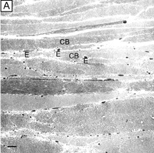
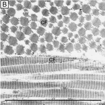
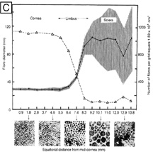
| Fig. 34. (A) Low (4,750×) and (B) high magnification (72,500×) electron micrographs of the human sclera in the region of the scleral stroma proper. Compare these to Figures 10A and B to see how much more irregular the collagen bundles are in the sclera and how much more variable the collagen fibril diameters and spaces are. CB, collagen bundle; E, elastin fibers; CF, collagen fibril. (C) Summary diagram comparing the collagen fibril diameters and densities in the cornea, limbus, and sclera. ([C] is from Borcherding MS, et al. Proteoglycans and collagen fibre organization in human corneoscleral tissue. Exp Eye Res 21:59, 1975.) |