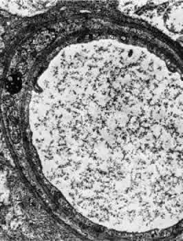
Fig. 5.
Electron micrograph of a human retinal venule. E, endothelial cell. The pericyte (P) contains abundant mitochondria and rough-surfaced endoplasmic reticulum. Pinocytotic vesicles are present, and the myofilaments are poorly developed.