|
|
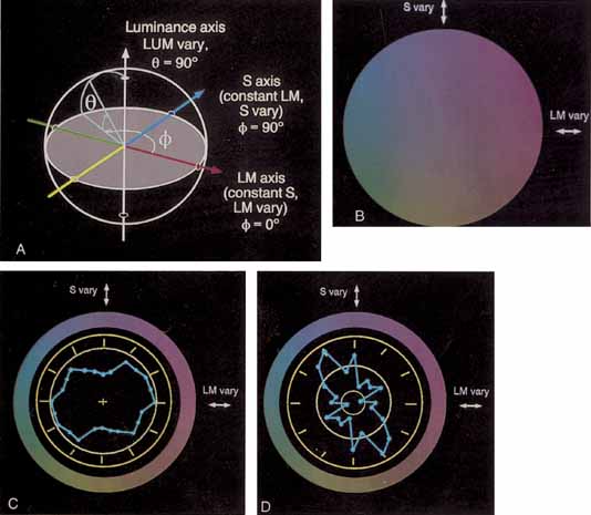
| Fig. 21 A: Three-dimensional color space for specification of cone stimulation. The three orthogonal axes in are constant S along which only L and M cone stimulation changes, constant LM along which only S cones are modulated (i.e., a tritanopic confusion axis), achromatic or luminance along which the sum stimulation of all three cone types changes, but not their relative excitations. Any modulation can be specified by the azimuth (ϕ) and elevation (Θ) of the vector joining the origin and a point on the surface of the sphere. The shaded area indicates the isoluminant plane through the achromatic origin. (Adapted from Derrington AM, Krauskopf J, Lennie P: Chromatic mechanisms in lateral geniculate nucleus of macaque. J Physiol Lond 357:241, 1984) B: Actual color variation in the isoluminant plane noted in A. C: Polar plot of the VEP responses from a patient with a history of central serous choroidopathy. The ratios of the amplitudes (affected eye to unaffected eye) are plotted as a function of direction in the isoluminant plane. The inner circle indicates a ratio of 1 (i.e., equivalent amplitudes in each eye). D: Same as in C except the difference in latencies between the eyes is plotted (inner circle = zero difference; second circle = 20-msec difference). |