|
|
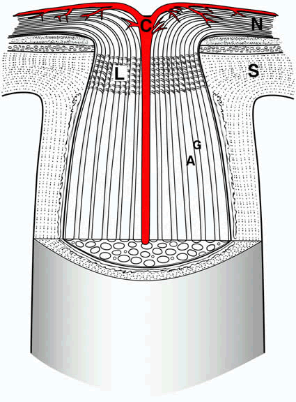
| Fig. 3. Cross-sectional view of the optic nerve head. For simplification, only the central retinal artery (C) is shown. Retinal ganglion cell axons arising in the nerve fiber layer (N) course in bundles (A) within the optic nerve, separated by glia (G). The lamina cribrosa (L) is contiguous to the sclera (S). |