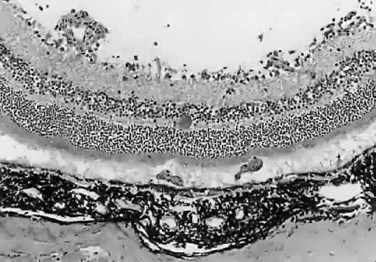
Fig. 3.
Light micrograph of a hamster eye after resolution of ocular toxoplasmosis. A large tissue cyst is seen in normalappearing retina (hematoxylin and eosin). (Courtesy of Barbara A. Nichols, PhD.)