|
|
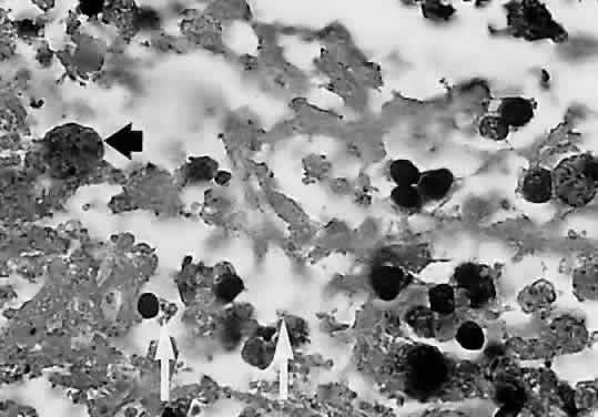
| Fig. 5. Light micrograph of a necrotic retinal lesion examined at the autopsy of a patient with AIDS and ocular toxoplasmosis. Tissue cysts (black arrow) and trophozoites (white arrows) are seen. There is little inflammatory material (hematoxylin and eosin). (Holland GN, Engstrom RE, Glasgow BJ et al: Ocular toxoplasmosis in patients with acquired immunodeficiency syndrome. Am J Ophthalmol 106:653, 1988. Copyright The Ophthalmic Publishing Company.) |