|
|
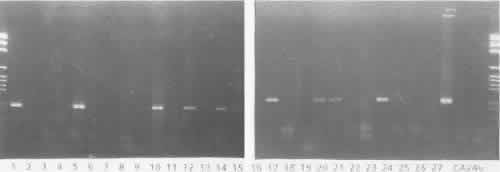
| Fig. 6. Polymerase chain reaction analysis of clinical conjunctival swab specimens deemed enterovirus tissue-culture negative using VP1 region primer (top right and left) and VP2 region primer (bottom right and left). Ethidium bromide-stained plates are shown. Lanes 1–27, clinical samples; lane 28, enterovirus 70 prototype virus (J670/71); lane 29, CA24v prototype virus (EH24/70); lane 30, negative control. mk represents the 1-kilobase pair ladder marker. The molecular weights of the polymerase chain reaction products are indicated to the side of the figures. Lanes 1, 5, 9, 10, 12, 14, 15, 17, 20, 21, and 24 show a positive signal in both primers, but lane 8 shows a positive signal only in the VP2 primer. |