|
|
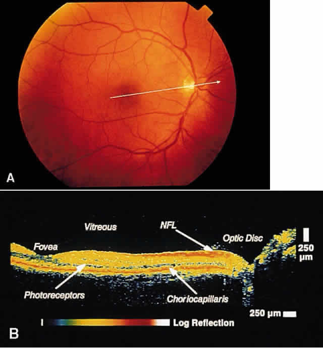
| Fig. 1. Color photograph of normal fundus. The white line indicates the area of retina and optic disc scanned on the corresponding OCT in B. B. OCT image through papillomacular bundle shown in A. The vitreoretinal interface, individual retinal layers, choriocapillaris, foveal contour, and optic disc are well delineated. |