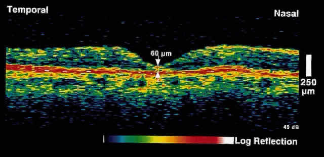
Fig. 12.
OCT image through the fovea of the same patient after macular hole surgery. Note the restoration of normal foveal anatomy.