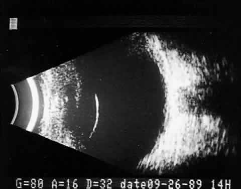
Fig. 2.
Contact B-scan. Cross-sections are presented horizontally. Normal globe, anteroposterior view with lens capsule seen toward the left of the display, optic nerve and orbital fat behind the globe seen toward the right.