|
|
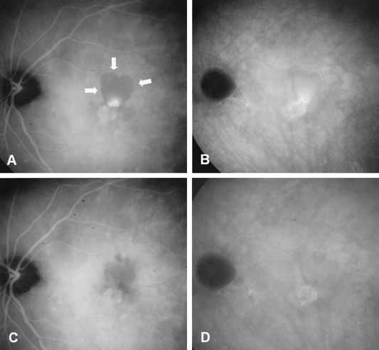
| Fig. 5 A, Mid-phase indocyanine green (ICG) angiogram shows an area of hyperfluorescence surrounded by an area of hypofluorescence (arrows) in a patient with polypoidal choroidal vasculopathy representing a focal area of active neovascularization surrounded by pigment epithelium detachment (PED). B, Late-phase ICG reveals hyperfluorescence of the vascular network. C, D, The patient was treated with photodynamic therapy using verteporfin. C, Mid-phase and late-phase ICG angiograms, which were performed 5 months after treatment, demonstrate resolution of fluid, flattened PED, and no active leakage. D, There is some late staining from the vascular network in the late-phase ICG angiogram. |