|
|
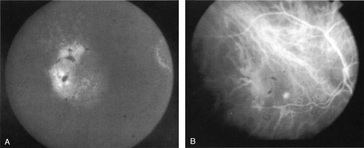
| Fig. 1 A. Late-phase fluorescein angiogram of a patient with exudative age-related macular degeneration demonstrating diffuse hyperfluorescence and staining of a serous retinal pigment epithelial detachment. No localized area of classic neovascularization was identified, and the patient was classified as having occult choroidal neovascularization. B. Mid-phase indocyanine green angiogram demonstrating a focal spot of hyperfluorescence presumed to represent a localized area of choroidal neovascularization. |