|
|
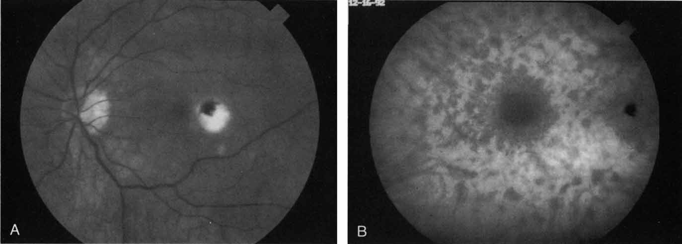
| Fig. 18 A. Clinical photograph of a patient with active inflammation associated with multifocal choroiditis. An atrophic scar from previous laser photocoagulation treatment is noted in the temporal macula. The patient reported visual disturbance, and an enlarged blind spot was noted on visual field testing. B. Late-phase indocyanine green study demonstrating a multitude of hypofluorescent spots that were larger than those seen with multiple evanescent white dot syndrome and more numerous and more extensively distributed than appreciated on clinical examination. Note that there is marked confluence of these lesions around the optic nerve, which may help to explain the enlarged blind spot noted on visual field testing. |