|
|
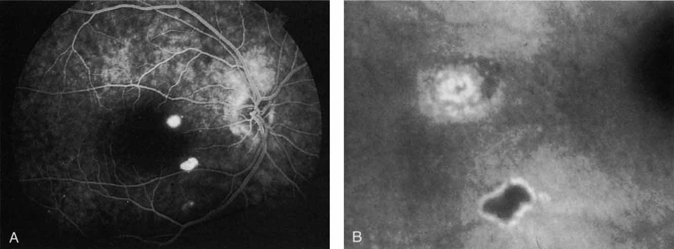
| Fig. 21 A. Late-phase fluorescein study demonstrating hyperfluorescent retinal pigment epithelial detachments in this patient with recent visual symptoms. B. High-magnification of the late-phase of the indocyanine green study, which shows the two pigment epithelial detachments that were hyperfluorescent on the fluorescein angiogram (A). The superior lesion is more active, demonstrating a central hypofluorescence surrounded by a ring of hyperfluorescence, characteristic of the pigment epithelial detachments in patients with central serous chorioretinopathy. |