|
|
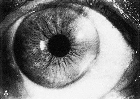
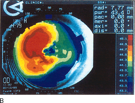
| Fig. 13. Terrien's marginal degeneration. A. Slit lamp photograph showing peripheral corneal thinning in the inferior and superior nasal quadrants of the right eye. Two areas with prominent peripheral white lines were present from the 1- to 3-o'clock and the 3:30- to 6-o'clock positions. A small area of normal cornea separates the abnormal area from the limbus. B. Topographic map (normalized scale) demonstrates flattening of the superior and inferior nasal peripheral cornea that corresponds to the area of thinning. This flattening compromises the visual axis and produces less than 1 diopter of cylinder, detected also by refraction and keratometry. Wilson S, Lin D, Klyce S et al: Terrien's marginal degeneration: Corneal topography. Refract Corneal Surg 6:15–20, 1990.) |